Chapter 2 : Pathological Slides
Topics covered in this snack-sized chapter:
When a patient has a biopsy or surgery, the surgeon often will remove diseased tissue for examination by the hospital's pathology department. The tissue is referred to as a “Tissue block”.
The pathologist will slice the tissue block into very thin layers that are placed on a glass slide and examined under a microscope.
This allows the pathologist to assist the surgeon in confirming a diagnosis of the diseased tissue.
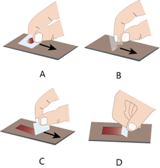
Semisolid aspirates are crush-smeared by flat pressure with cover slip or glass slide.
Fluid or blood droplet is collected along edge of spreader.
The fluid is pulled along the slide to create a thin film.
This film is called smear.
Steps of Making a Pathological Microscopic Slide arrow_upward
Making a microscope slide starts with the fixation of your biopsy or necropsy specimen.
- The majority of samples are fixed in 10% neutral buffered formalin.
- Ratio of sample volume to formalin volume should be at least 1:10.
- Once the tissues are properly fixed, it is trimmed to fit into a tissue cassette.
Tissue cassettes come in different sizes and colors.
- For standard cassettes, the tissue should be about “2 nickels” thick.
- Tissues in the cassettes are stored in formalin and transferred to the histology lab.
- At the “histo lab” the cassettes are processed. Processing removes the formalin and water from the tissues and replaces it with wax.
The “wax” tissues are then embedded in wax, and the result is called a “block”.
A microtome is used to slice sections off the block in the form of a “ribbon.” The microtome can be set to cut at different thicknesses - for instance, 4 microns, or 7 microns.
The delicate ribbon is carefully transferred to a warm water bath.
The ribbon floats on the surface and a slide can be placed under the water to lift the section up.
The slide is left to dry, and then it is placed in a dry oven which will gently melt the excess parafin from the section, leaving the section of tissue intact.
After removal of the excess wax, the slides can be stained with hematoxilin and eosin.
This can be done by hand, or by an automated stainer.
Special stains, such as trichrome stains and Geimsa’s, are handled separately.
After slides are stained, the final step is to cover slip them.
This can be done by hand or by an automated coverslipper and see in microscope.