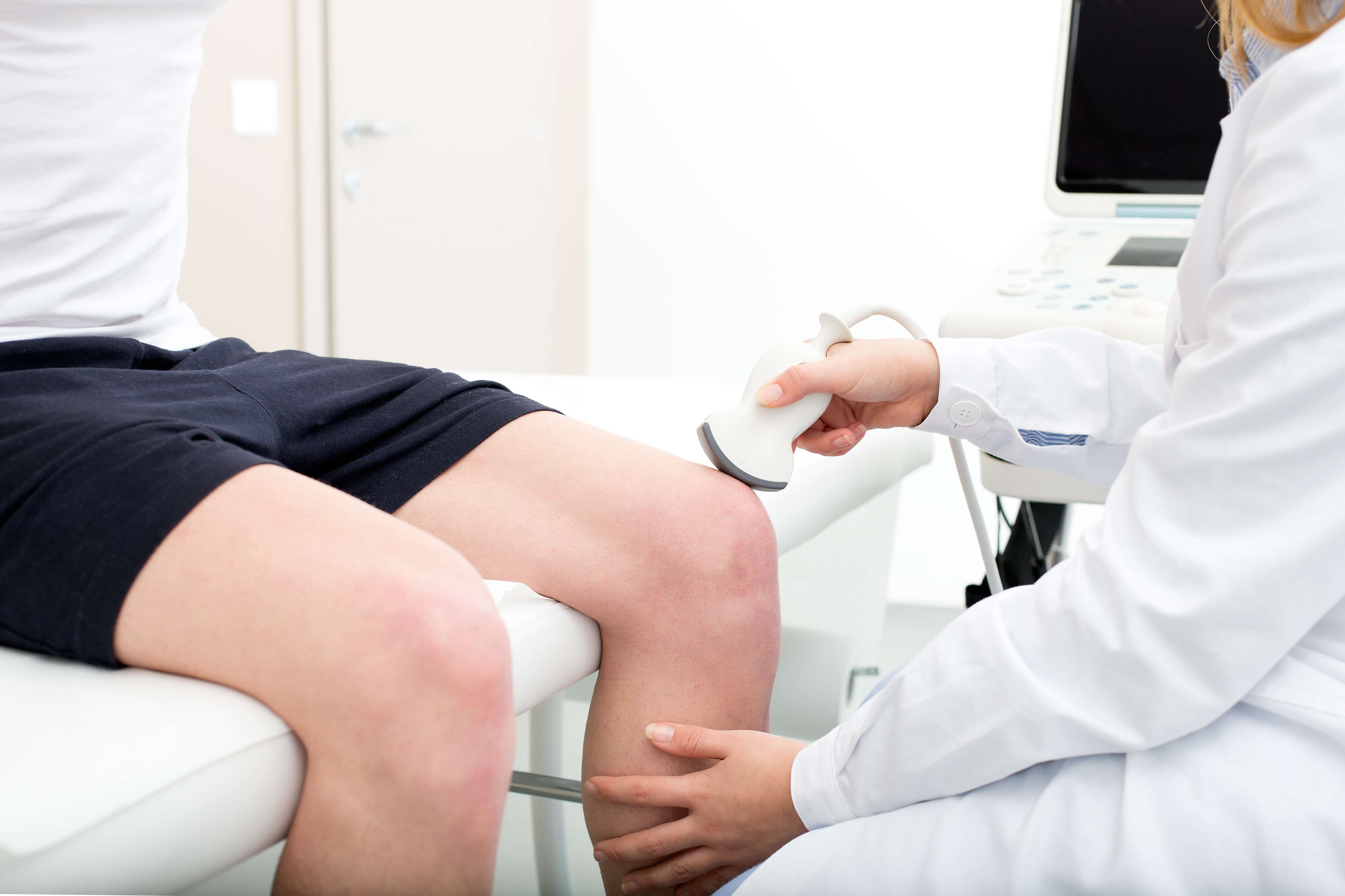
Manual muscle testing is a common method used in physical therapy to assess muscle strength by evaluating the ability of a muscle or muscle group to generate force against resistance. This technique involves the therapist applying resistance while the patient performs specific movements, allowing for the determination of muscle strength and any potential weaknesses or imbalances. By systematically testing different muscle groups, therapists can identify areas of weakness and tailor treatment plans accordingly.
In manual muscle testing, different grades are used to classify muscle strength based on the patient's ability to resist the applied resistance. The grades range from 0 to 5, with 0 indicating no muscle contraction, 1 representing a trace contraction, 2 indicating movement with gravity eliminated, 3 representing movement against gravity, 4 indicating movement against resistance, and 5 representing normal muscle strength. These grades help therapists quantify and track changes in muscle strength over time.
Pediatric PT Approved Foam Roller Exercises & Activities! The post Foam Roller Appreciation appeared first on DINOSAUR PHYSICAL THERAPY.
Posted by on 2024-03-28
Pediatric PT Approved Early Riding Toys! The post Our Favorite Early Riding Toys appeared first on DINOSAUR PHYSICAL THERAPY.
Posted by on 2024-03-01
Alternative Seating Options for Children! The post Alternative Seating Options appeared first on DINOSAUR PHYSICAL THERAPY.
Posted by on 2024-02-12
Pediatric PT Approved Cold Weather Shoe Recommendations! The post Shoe Recommendations for Children: Cold Weather Edition appeared first on DINOSAUR PHYSICAL THERAPY.
Posted by on 2024-01-31
Manual muscle testing can be a valuable tool in monitoring progress in muscle rehabilitation programs. By regularly assessing muscle strength using this method, therapists can track improvements or declines in strength, adjust treatment plans as needed, and set realistic goals for the patient's recovery. This objective measurement allows for a more targeted and effective approach to rehabilitation.
Despite its benefits, manual muscle testing has limitations in assessing muscle strength. Factors such as patient effort, therapist experience, and variability in resistance application can influence the results. Additionally, the subjective nature of grading muscle strength can lead to inconsistencies between different therapists. It is important for therapists to be aware of these limitations and use manual muscle testing in conjunction with other assessment tools for a comprehensive evaluation.
The position of the joint during manual muscle testing can significantly impact the results obtained. The angle at which the muscle is tested can affect the leverage and mechanical advantage, leading to variations in strength measurements. It is crucial for therapists to standardize the testing positions to ensure accurate and reliable results. Consistency in joint positioning helps minimize confounding variables and allows for more meaningful comparisons over time.
When performing manual muscle testing, there are common errors that therapists should avoid to ensure accurate results. Some of these errors include inadequate stabilization of the patient or the therapist, inconsistent application of resistance, improper positioning of the patient or the therapist, and failure to communicate effectively with the patient. By being mindful of these potential pitfalls and following standardized protocols, therapists can minimize errors and obtain reliable muscle strength assessments.
Manual muscle testing can help differentiate between muscle weakness and neurological deficits by assessing the quality of muscle contractions. In cases of muscle weakness, the patient may exhibit reduced strength in specific muscle groups, while maintaining normal coordination and sensation. On the other hand, neurological deficits may present as weakness accompanied by other signs such as altered reflexes, muscle tone, or coordination. By carefully evaluating the patient's response to resistance during manual muscle testing, therapists can identify the underlying cause of the impairment and tailor treatment accordingly.
ROM testing plays a crucial role in post-surgical rehabilitation by assessing the range of motion in the affected joint or muscle. This testing helps physical therapists and healthcare professionals monitor the progress of the patient's recovery, identify any limitations or restrictions in movement, and determine the effectiveness of the rehabilitation program. By measuring the flexibility, strength, and mobility of the injured area, ROM testing allows for the development of personalized treatment plans that focus on improving functional abilities and reducing pain. Additionally, ROM testing can help prevent complications such as muscle stiffness, joint contractures, and scar tissue formation, ultimately promoting a faster and more successful recovery process for the patient.
ROM testing can be seamlessly integrated into a comprehensive physical assessment by incorporating a variety of specific movements and measurements to evaluate the flexibility and range of motion of different joints in the body. This can include assessing active and passive ROM, using goniometers to quantify joint angles, and performing special tests to identify any limitations or abnormalities. By incorporating ROM testing into the assessment process, healthcare professionals can gather valuable information about a patient's musculoskeletal health, identify potential areas of concern, and develop targeted treatment plans to improve overall function and mobility. Additionally, integrating ROM testing into a comprehensive physical assessment allows for a more thorough evaluation of a patient's physical capabilities and can help track progress over time.
Limited elbow extension range of motion (ROM) can be caused by a variety of factors, including joint stiffness, muscle tightness, ligamentous restrictions, joint effusion, osteoarthritis, bursitis, tendonitis, nerve compression, scar tissue formation, and previous elbow injuries. These conditions can lead to decreased flexibility, pain, swelling, and difficulty fully extending the elbow. Physical therapy, stretching exercises, manual therapy, joint mobilizations, and modalities such as heat and ice therapy can help improve elbow extension ROM and alleviate symptoms associated with these underlying causes. It is important to consult with a healthcare professional for an accurate diagnosis and appropriate treatment plan to address the specific issue causing limited elbow extension ROM.
When interpreting ROM testing results for spinal flexion, healthcare professionals typically look at the range of motion measurements taken during the assessment. These measurements provide valuable information about the flexibility and mobility of the spine in terms of forward bending. By analyzing the data collected, practitioners can determine if there are any restrictions, limitations, or abnormalities present in the spinal flexion movement. Additionally, they may compare the results to established norms or previous assessments to track progress or identify any changes over time. Overall, interpreting ROM testing results for spinal flexion involves a comprehensive analysis of the quantitative data to inform clinical decision-making and treatment planning.
Common range of motion deficits seen in postoperative knee patients include limited flexion and extension, decreased joint mobility, stiffness, and difficulty with activities of daily living. These deficits can be caused by factors such as scar tissue formation, muscle weakness, pain, swelling, and inflammation. Physical therapy interventions, such as stretching exercises, manual therapy, and modalities like heat and ice, are often used to address these deficits and improve knee function. It is important for healthcare providers to closely monitor and address ROM deficits in postoperative knee patients to optimize outcomes and prevent long-term complications.