
The formation of calcium deposits in the tendons is a key factor in the development of calcific tendinitis. These deposits can accumulate over time due to various factors such as repetitive stress or injury to the tendon. The presence of these deposits can lead to inflammation and irritation in the surrounding tissues, causing pain and discomfort in the affected area. Additionally, the calcium deposits can disrupt the normal structure and function of the tendon, further exacerbating the symptoms of calcific tendinitis.
Inflammatory responses play a significant role in the progression of calcific tendinitis. When calcium deposits form in the tendons, the body's immune system may perceive them as foreign substances and mount an inflammatory response to try to eliminate them. This inflammatory process can lead to swelling, redness, and pain in the affected area. In some cases, the inflammation can become chronic, contributing to the persistence of symptoms in individuals with calcific tendinitis.
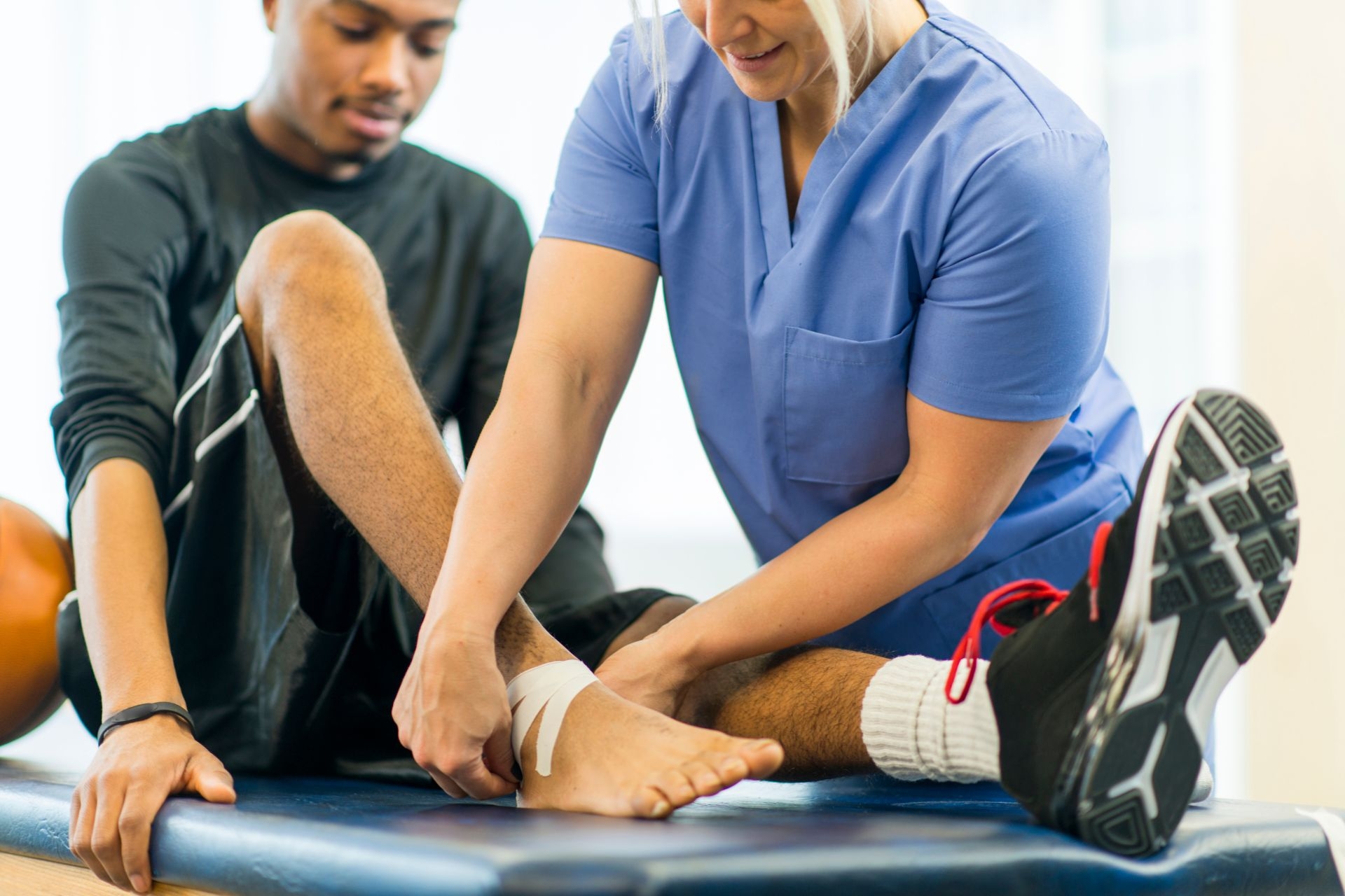
Core strength training is an important part of physical therapy. The muscles in your core help in anchoring your center of gravity, which gives you the ability to balance yourself. Whether you’re sitting, standing, or running, your core muscles play an integral role in keeping you balanced. A weak core... The post Improve Your Core Strength Through Your Balance! appeared first on APEX Physical Therapy.

Posted by on 2023-11-10
The presence of calcific deposits in the tendons can significantly affect the range of motion in individuals with calcific tendinitis. The deposits can cause stiffness and limited flexibility in the affected tendon, making it difficult for individuals to move the affected joint or muscle. This restriction in range of motion can further contribute to pain and discomfort, impacting the individual's ability to perform daily activities or participate in physical exercise.

Common symptoms experienced by individuals with calcific tendinitis include pain, swelling, tenderness, and stiffness in the affected tendon. The pain may worsen with movement or pressure on the tendon, and individuals may also experience difficulty performing certain activities that involve the affected joint or muscle. In some cases, the symptoms of calcific tendinitis may come and go, with periods of flare-ups followed by periods of relative relief.
Calcific tendinitis is typically diagnosed through a combination of physical examination, medical history review, and imaging tests. X-rays are commonly used to visualize the presence of calcium deposits in the affected tendon. Ultrasound or MRI scans may also be used to assess the extent of the calcific deposits and evaluate the condition of the surrounding tissues. These imaging techniques help healthcare providers confirm the diagnosis of calcific tendinitis and develop an appropriate treatment plan.
Treatment options for calcific tendinitis may include conservative measures such as rest, ice, physical therapy, and nonsteroidal anti-inflammatory drugs (NSAIDs) to manage pain and inflammation. In some cases, corticosteroid injections or extracorporeal shock wave therapy may be recommended to help break down the calcium deposits and promote healing in the affected tendon. Surgical intervention may be considered for severe cases of calcific tendinitis that do not respond to conservative treatments. The effectiveness of these treatment options may vary depending on the individual's specific condition and response to therapy.
There are several risk factors and underlying conditions that may increase the likelihood of developing calcific tendinitis. These include age, as the condition is more common in individuals over the age of 40, and gender, as women are more likely to be affected than men. Other risk factors may include a history of tendon injuries or overuse, certain medical conditions such as diabetes or thyroid disorders, and genetic predisposition. Understanding these risk factors can help healthcare providers identify individuals who may be at higher risk for developing calcific tendinitis and implement preventive measures to reduce the likelihood of occurrence.

Orthopedic physical therapy takes a comprehensive approach to rehabilitating individuals with iliotibial band syndrome, focusing on addressing the underlying biomechanical issues that contribute to the condition. Treatment may include targeted exercises to strengthen the hip abductors, gluteal muscles, and core stabilizers to improve alignment and reduce strain on the iliotibial band. Manual therapy techniques such as soft tissue mobilization and myofascial release may also be used to alleviate tightness and improve flexibility in the affected area. Additionally, gait analysis and running mechanics assessment may be conducted to identify and correct any faulty movement patterns that could be exacerbating the syndrome. By addressing these factors, orthopedic physical therapy aims to not only alleviate symptoms but also prevent future occurrences of iliotibial band syndrome.
Orthopedic physical therapy plays a crucial role in addressing the rehabilitation needs of individuals with chronic lower back pain by focusing on improving strength, flexibility, and mobility in the affected area. Therapists utilize a variety of techniques such as manual therapy, therapeutic exercises, and modalities like heat and ice to alleviate pain and improve function. Additionally, education on proper body mechanics, posture, and ergonomics is provided to prevent further injury and promote long-term relief. By addressing the underlying musculoskeletal imbalances and dysfunctions contributing to the pain, orthopedic physical therapy helps individuals regain function and quality of life. The personalized treatment plans are designed to target specific areas of weakness or tightness, promoting overall stability and resilience in the lower back region. Through a comprehensive approach that includes strengthening core muscles, improving flexibility in surrounding tissues, and addressing any biomechanical issues, orthopedic physical therapy effectively addresses the unique rehabilitation needs of individuals with chronic lower back pain.
Kinesiology taping in orthopedic physical therapy offers several potential benefits for patients. The application of kinesiology tape can help improve circulation, reduce inflammation, and provide support to injured or weak muscles and joints. This can lead to decreased pain, improved range of motion, and enhanced proprioception. Additionally, kinesiology taping can help facilitate proper movement patterns and muscle activation, aiding in the rehabilitation process. The tape's elastic properties allow for full range of motion while still providing support, making it a versatile tool in orthopedic physical therapy. Overall, kinesiology taping can be a valuable adjunct to traditional treatment methods in helping patients recover from orthopedic injuries and conditions.
Orthopedic physical therapy can be beneficial in enhancing joint mobility for individuals diagnosed with tarsal tunnel syndrome. By focusing on specific exercises and techniques tailored to the affected area, physical therapists can help improve range of motion, reduce pain, and increase overall function in the foot and ankle joints. Through targeted interventions such as stretching, strengthening, and manual therapy, patients with tarsal tunnel syndrome can experience improvements in joint flexibility, stability, and proprioception. Additionally, orthopedic physical therapy may also address any underlying biomechanical issues contributing to the condition, further enhancing joint mobility and overall quality of life for individuals with tarsal tunnel syndrome.
In orthopedic physical therapy for patients with patellar tendinopathy, recommended modifications for plyometric exercises may include reducing the intensity and volume of the exercises, focusing on eccentric loading, incorporating isometric exercises, and utilizing proper technique and form. It is important to avoid high-impact activities that place excessive stress on the patellar tendon, such as deep squats or jumping exercises. Instead, therapists may prescribe exercises that target the quadriceps, hamstrings, and glutes while minimizing strain on the patellar tendon. Additionally, implementing a gradual progression of plyometric exercises and monitoring for any signs of pain or discomfort is crucial in managing patellar tendinopathy effectively. By tailoring plyometric exercises to the specific needs and limitations of patients with patellar tendinopathy, physical therapists can help improve strength, function, and overall outcomes in rehabilitation.
Orthopedic physical therapy can be beneficial in improving ankle dorsiflexion range of motion by utilizing a variety of techniques such as manual therapy, stretching exercises, strengthening exercises, and proprioceptive training. By targeting the muscles, tendons, ligaments, and joints surrounding the ankle joint, physical therapists can help increase flexibility, reduce stiffness, and improve overall function. Additionally, modalities such as ultrasound, electrical stimulation, and heat/cold therapy may be used to further enhance the effects of treatment. Through a comprehensive rehabilitation program tailored to the individual's specific needs, orthopedic physical therapy can effectively address limitations in ankle dorsiflexion range of motion and promote optimal recovery.