
Glenohumeral joint dislocation commonly occurs due to traumatic events such as falls, sports injuries, or motor vehicle accidents. The forceful impact on the shoulder can cause the humerus bone to pop out of the glenoid socket, leading to dislocation. Additionally, repetitive overhead movements or instability in the shoulder joint can also contribute to the risk of dislocation.
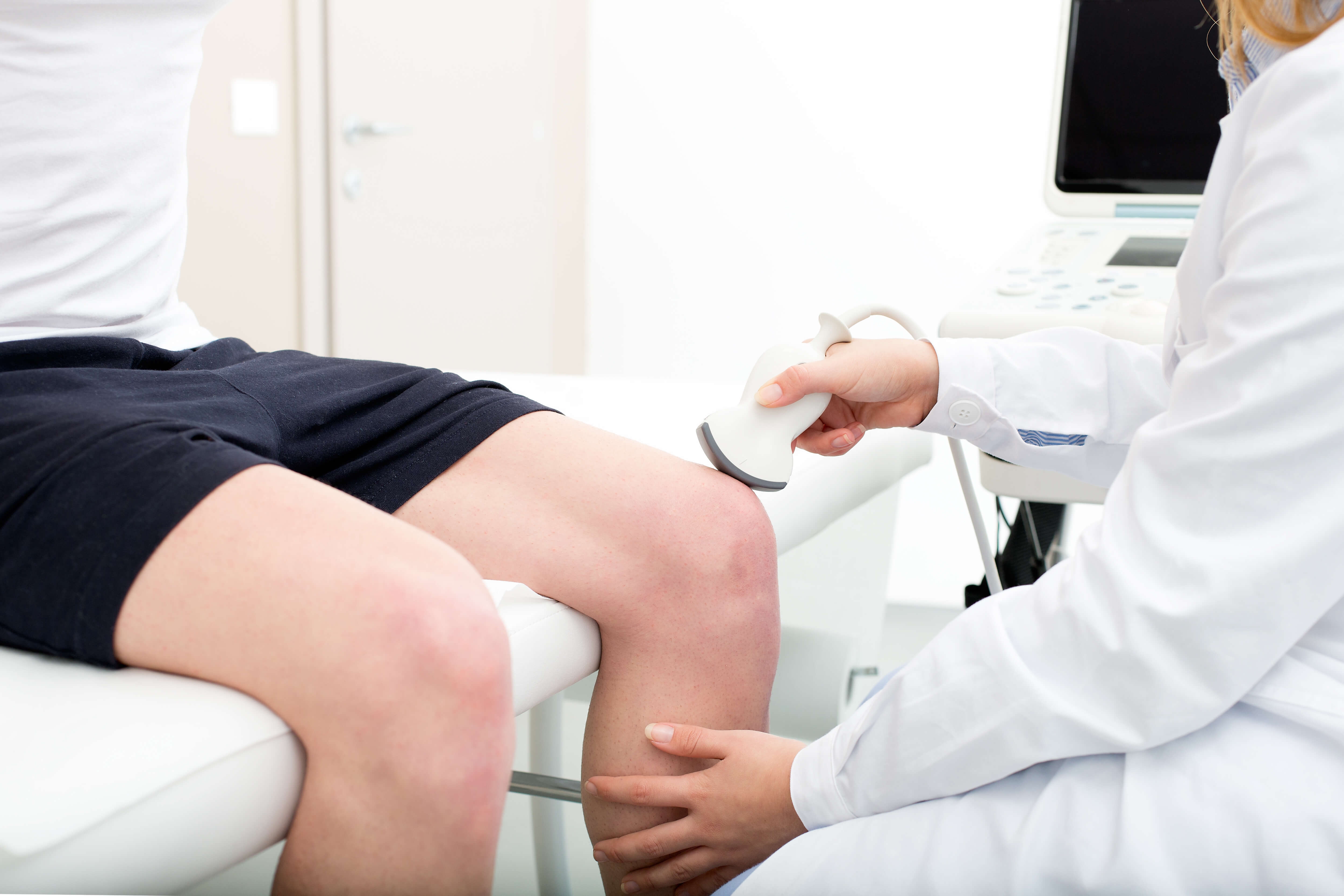
Healthcare professionals diagnose a glenohumeral joint dislocation through a physical examination, where they assess the range of motion, stability, and tenderness in the shoulder. X-rays are often used to confirm the dislocation and determine the extent of the injury. In some cases, an MRI or CT scan may be recommended to evaluate any associated soft tissue damage.
Core strength training is an important part of physical therapy. The muscles in your core help in anchoring your center of gravity, which gives you the ability to balance yourself. Whether you’re sitting, standing, or running, your core muscles play an integral role in keeping you balanced. A weak core... The post Improve Your Core Strength Through Your Balance! appeared first on APEX Physical Therapy.
Posted by on 2023-11-10
Individuals with a glenohumeral joint dislocation typically experience intense pain, swelling, and bruising in the shoulder area. They may also have difficulty moving the affected arm and notice a visible deformity in the shoulder joint. Numbness or tingling sensations in the arm or hand can also occur due to nerve compression.
There are different types of glenohumeral joint dislocations, including anterior dislocation (most common), posterior dislocation, inferior dislocation, and multidirectional dislocation. Anterior dislocation occurs when the humerus moves forward out of the socket, while posterior dislocation involves the humerus moving backward. Inferior dislocation occurs when the humerus moves downward, and multidirectional dislocation involves instability in multiple directions.
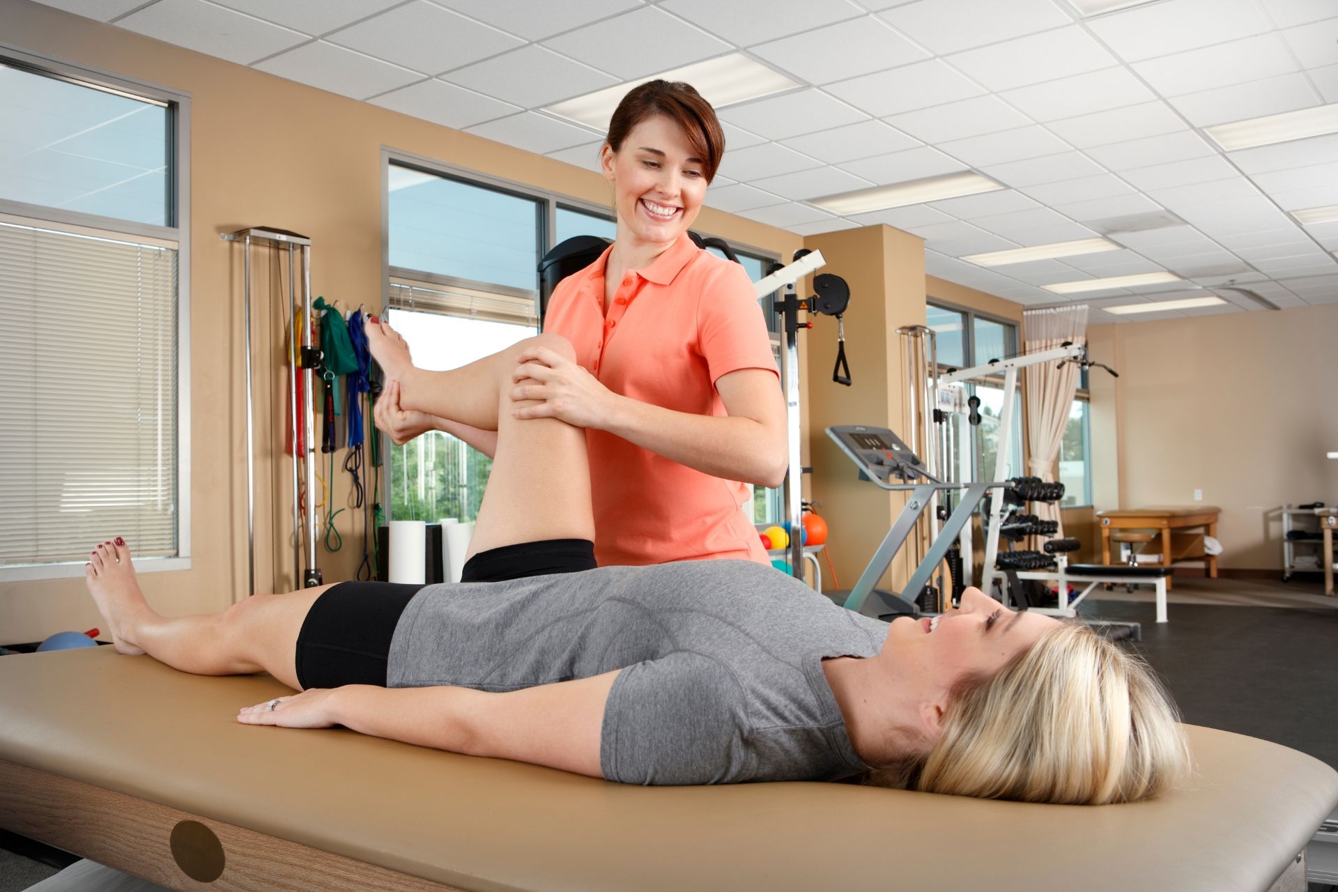
Non-surgical treatment for a glenohumeral joint dislocation often involves closed reduction, where the healthcare provider manipulates the shoulder joint to realign the bones. Following reduction, immobilization with a sling, physical therapy, and strengthening exercises are typically recommended. In cases of recurrent dislocations or severe damage, surgical intervention such as arthroscopic stabilization or open surgery may be necessary.
Specific risk factors that increase the likelihood of experiencing a glenohumeral joint dislocation include participation in contact sports, such as football or rugby, where there is a higher risk of traumatic shoulder injuries. Individuals with a history of shoulder instability, lax ligaments, or previous dislocations are also more prone to experiencing recurrent dislocations.
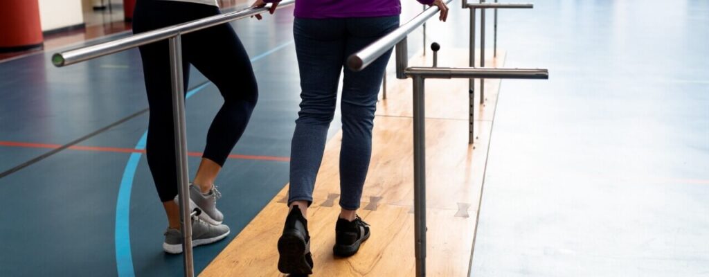
The typical recovery time for individuals who have suffered from a glenohumeral joint dislocation varies depending on the severity of the injury and the chosen treatment approach. In general, non-surgically treated dislocations may take several weeks to months for full recovery, including rehabilitation to regain strength and range of motion. Surgical interventions may require a longer recovery period, with rehabilitation playing a crucial role in restoring shoulder function. It is essential to follow the healthcare provider's recommendations for a successful recovery and to prevent future dislocations.
Orthopedic physical therapy can be beneficial in enhancing joint mobility in individuals diagnosed with ankylosing spondylitis, a chronic inflammatory condition that primarily affects the spine and sacroiliac joints. Through targeted exercises, manual therapy techniques, and modalities such as heat and cold therapy, physical therapists can help improve range of motion, flexibility, and overall function in patients with ankylosing spondylitis. By focusing on strengthening muscles surrounding the affected joints, promoting proper posture, and addressing any postural abnormalities, orthopedic physical therapy can play a crucial role in managing symptoms and preventing further joint stiffness and immobility. Additionally, education on proper body mechanics and lifestyle modifications may also be provided to empower individuals with ankylosing spondylitis to better manage their condition and improve their quality of life.
Orthopedic physical therapy can be a beneficial treatment option for athletes suffering from patellar tendinitis. By focusing on specific exercises and techniques designed to strengthen the muscles surrounding the knee, improve flexibility, and address any biomechanical issues contributing to the condition, orthopedic physical therapy can help athletes recover and return to their sport. Additionally, modalities such as ultrasound, electrical stimulation, and manual therapy may be utilized to reduce pain and inflammation in the affected area. By working closely with a physical therapist who specializes in orthopedic care, athletes with patellar tendinitis can improve their symptoms and prevent future injury.
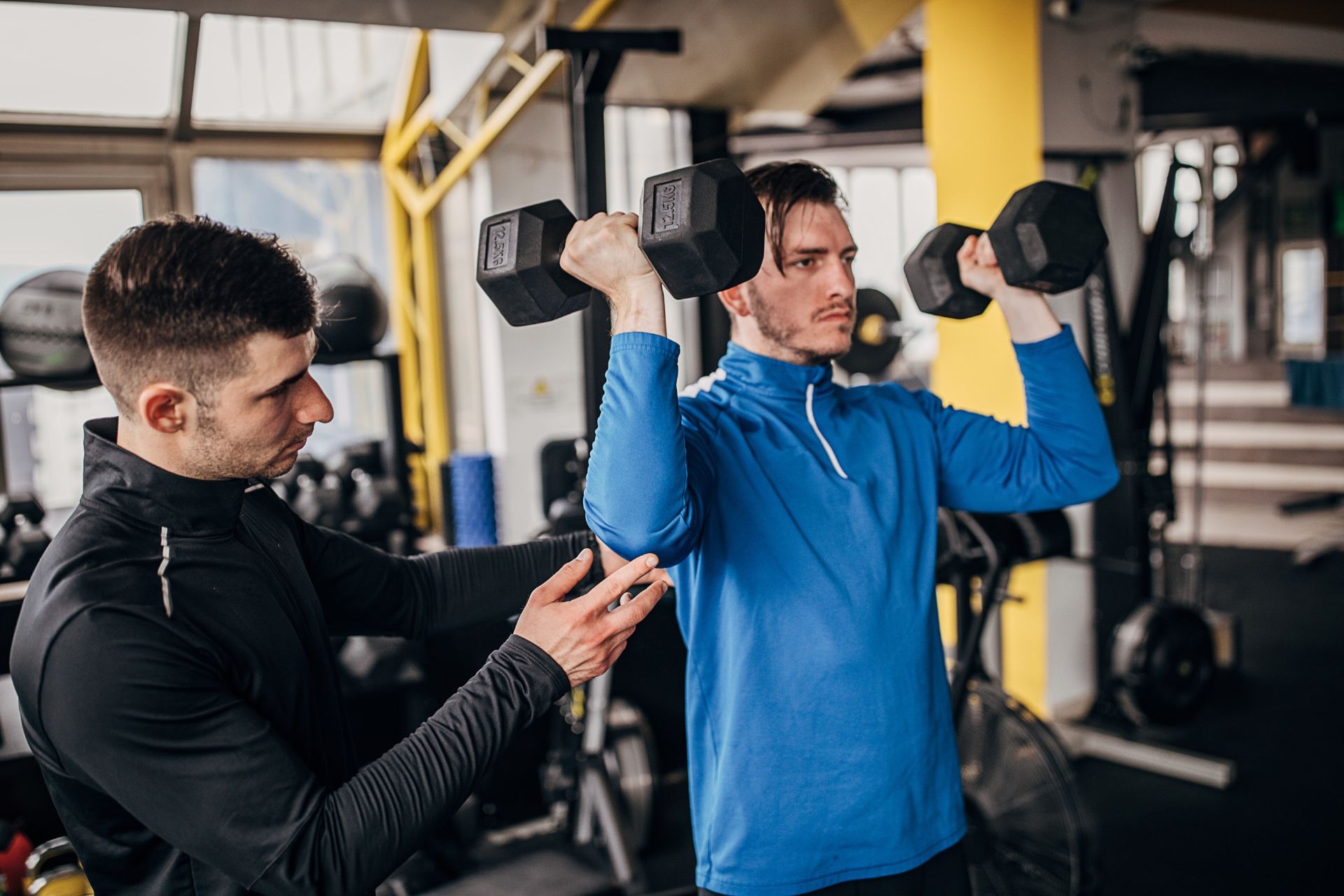
Orthopedic physical therapy plays a crucial role in preventing common sports injuries by focusing on specific strategies tailored to each individual's needs. Some effective approaches include implementing proper warm-up and cool-down routines, incorporating strength and flexibility exercises, utilizing proper technique and form during sports activities, addressing muscle imbalances and weaknesses, promoting proper nutrition and hydration, emphasizing rest and recovery periods, and utilizing appropriate protective gear and equipment. By addressing these key components through orthopedic physical therapy, individuals can reduce their risk of sustaining common sports injuries such as sprains, strains, fractures, and overuse injuries. Additionally, orthopedic physical therapy can help improve overall performance and enhance athletic abilities, leading to a more successful and injury-free sports experience.
Orthopedic physical therapy approaches muscle imbalances in individuals with lateral pelvic tilt by focusing on targeted exercises to strengthen weak muscles and stretch tight muscles. This may involve exercises to improve core stability, hip flexibility, and pelvic alignment. By addressing specific muscle groups such as the hip abductors, adductors, and rotators, physical therapists can help restore balance and alignment in the pelvis. Additionally, manual therapy techniques such as soft tissue mobilization and joint mobilization may be used to address any restrictions or tightness contributing to the lateral pelvic tilt. Through a comprehensive treatment plan tailored to the individual's needs, orthopedic physical therapy aims to correct muscle imbalances and improve overall function and movement patterns.
Orthopedic physical therapy can play a crucial role in enhancing joint stability in individuals diagnosed with Ehlers-Danlos syndrome (EDS). By focusing on strengthening muscles surrounding the affected joints, improving proprioception, and enhancing overall body awareness, physical therapists can help patients with EDS better control their joint movements and reduce the risk of dislocations or subluxations. Additionally, targeted exercises aimed at improving posture, balance, and coordination can further contribute to enhancing joint stability in individuals with EDS. Through a comprehensive rehabilitation program tailored to the specific needs of each patient, orthopedic physical therapy can effectively address joint instability issues commonly associated with Ehlers-Danlos syndrome.
Orthopedic physical therapy for individuals with lumbar hyperlordosis focuses on addressing muscle tightness and imbalances through a combination of targeted exercises, manual therapy techniques, and postural education. Specific exercises such as pelvic tilts, hip flexor stretches, and core strengthening exercises are utilized to help correct muscle imbalances and improve flexibility in the hip flexors, hamstrings, and lower back muscles. Manual therapy techniques such as soft tissue mobilization and myofascial release may also be used to release tension in tight muscles and improve overall range of motion. Additionally, postural education plays a crucial role in helping individuals with lumbar hyperlordosis maintain proper alignment and prevent further muscle imbalances. By addressing these issues through a comprehensive orthopedic physical therapy program, individuals with lumbar hyperlordosis can experience improved mobility, reduced pain, and better overall function.
In orthopedic physical therapy, recommended exercises for strengthening the muscles surrounding the knee joint may include leg presses, squats, lunges, step-ups, hamstring curls, calf raises, and leg extensions. These exercises target the quadriceps, hamstrings, calves, and glutes, which are essential for providing stability and support to the knee joint. Additionally, incorporating balance and stability exercises such as single-leg stands, heel raises, and side leg lifts can help improve proprioception and prevent future injuries. It is important to gradually increase the intensity and resistance of these exercises to continue challenging the muscles and promoting strength gains in the knee joint. Proper form and technique should always be emphasized to prevent any further strain or injury to the knee.