
Intermetatarsal neuroma is a condition that affects the metatarsal bones in the foot by causing inflammation and compression of the nerves between the metatarsal heads. This can lead to pain, numbness, and tingling sensations in the affected area, typically between the third and fourth metatarsal bones.
Common symptoms associated with intermetatarsal neuroma include sharp or burning pain in the ball of the foot, a feeling of a pebble or lump under the foot, numbness or tingling in the toes, and discomfort that worsens with walking or wearing tight shoes. These symptoms can significantly impact an individual's ability to walk and engage in daily activities.
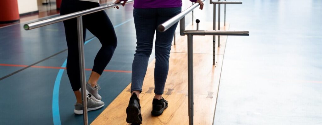
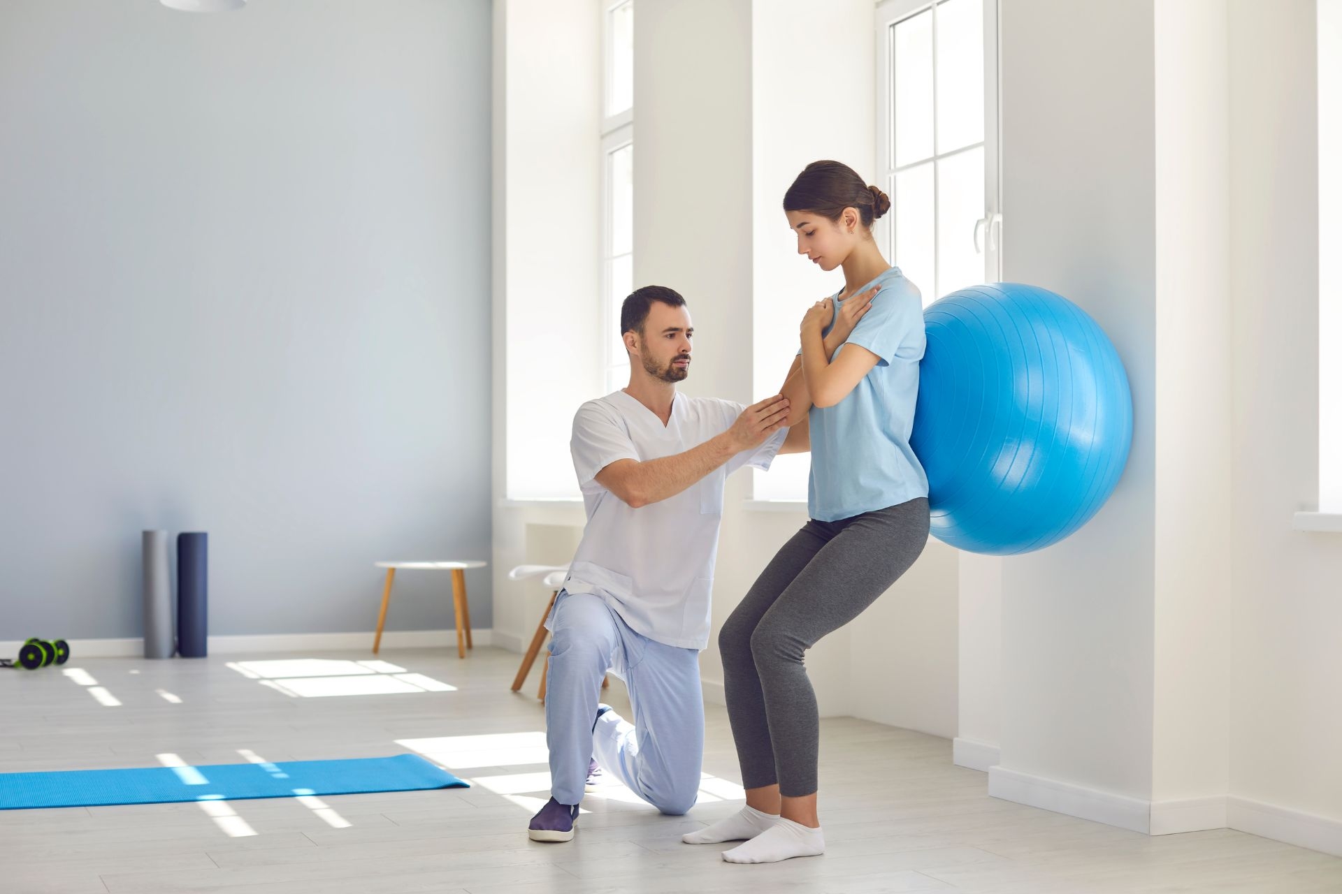
Core strength training is an important part of physical therapy. The muscles in your core help in anchoring your center of gravity, which gives you the ability to balance yourself. Whether you’re sitting, standing, or running, your core muscles play an integral role in keeping you balanced. A weak core... The post Improve Your Core Strength Through Your Balance! appeared first on APEX Physical Therapy.
Posted by on 2023-11-10
Healthcare professionals diagnose intermetatarsal neuroma through a combination of physical examination, medical history review, and imaging tests such as X-rays or MRI scans. The physical exam may involve applying pressure to the affected area to reproduce symptoms and assess the range of motion in the foot.

Treatment options for intermetatarsal neuroma may include conservative measures such as wearing supportive footwear, using orthotic inserts, taking anti-inflammatory medications, and receiving corticosteroid injections to reduce inflammation and pain. In more severe cases, surgical intervention to release the compressed nerve may be necessary.
Intermetatarsal neuroma can be prevented or managed through lifestyle changes such as wearing properly fitting shoes with adequate support, avoiding high heels or narrow-toed shoes that can exacerbate symptoms, maintaining a healthy weight to reduce pressure on the feet, and performing stretching exercises to improve foot flexibility.

Specific risk factors that increase the likelihood of developing intermetatarsal neuroma include wearing high heels or tight shoes regularly, participating in high-impact activities that put stress on the feet, having certain foot deformities such as flat feet or high arches, and experiencing repetitive trauma to the foot from sports or occupational activities.
Intermetatarsal neuroma differs from other foot conditions such as plantar fasciitis or bunions in terms of the underlying causes and symptoms. While plantar fasciitis involves inflammation of the tissue connecting the heel bone to the toes, intermetatarsal neuroma specifically affects the nerves between the metatarsal bones. Bunions, on the other hand, are bony protrusions that form at the base of the big toe, causing pain and deformity in the joint. Each condition requires a tailored treatment approach based on its unique characteristics.
Exercises that are recommended for improving hip internal rotation strength include clamshells, hip internal rotation with resistance bands, seated hip internal rotation, hip internal rotation with a stability ball, and hip internal rotation with a foam roller. These exercises target the muscles responsible for hip internal rotation, such as the gluteus medius, gluteus minimus, and tensor fasciae latae. Strengthening these muscles can help improve hip stability, reduce the risk of injury, and enhance overall lower body strength and function. It is important to perform these exercises with proper form and gradually increase resistance to continue challenging the muscles and promoting strength gains in hip internal rotation.
Orthopedic physical therapy approaches osteoarthritis and rheumatoid arthritis differently due to the distinct nature of these conditions. In treating osteoarthritis, physical therapists focus on improving joint function, reducing pain, and increasing mobility through exercises that strengthen the muscles surrounding the affected joint, as well as manual therapy techniques such as joint mobilization. In contrast, when treating rheumatoid arthritis, physical therapists aim to reduce inflammation, preserve joint integrity, and improve overall function through a combination of gentle exercises, modalities like heat and cold therapy, and education on joint protection techniques. Additionally, in rheumatoid arthritis, the emphasis may be on preventing deformities and maintaining range of motion in the affected joints.
In orthopedic physical therapy, specific exercises are often tailored for rehabilitating a rotator cuff tear. These exercises typically focus on strengthening the muscles surrounding the shoulder joint, such as the supraspinatus, infraspinatus, teres minor, and subscapularis. Common exercises may include external rotation exercises using resistance bands, internal rotation exercises with dumbbells, scapular stabilization exercises, and shoulder abduction exercises. Additionally, stretching exercises to improve flexibility in the shoulder joint and improve range of motion may also be incorporated into the rehabilitation program. It is important for individuals undergoing rotator cuff tear rehabilitation to work closely with a physical therapist to ensure proper form and progression of exercises to promote healing and prevent further injury.
Orthopedic physical therapy approaches muscle imbalances in individuals with anterior pelvic tilt by focusing on strengthening the weak muscles and stretching the tight muscles associated with this postural deviation. Specific exercises targeting the hip flexors, hamstrings, glutes, and core muscles are commonly prescribed to address the imbalance between the anterior and posterior muscle groups. Additionally, manual therapy techniques such as myofascial release and joint mobilizations may be used to improve muscle flexibility and joint alignment. Education on proper body mechanics and posture correction is also emphasized to prevent further exacerbation of the pelvic tilt. By addressing these muscle imbalances through a comprehensive treatment plan, orthopedic physical therapy aims to restore optimal alignment and function in individuals with anterior pelvic tilt.
Aquatic therapy in orthopedic physical rehabilitation offers numerous potential benefits due to the unique properties of water. The buoyancy of water reduces the impact on joints, allowing for low-impact exercises that can improve range of motion, strength, and flexibility. The resistance of water provides a gentle yet effective way to strengthen muscles without causing excessive strain. Additionally, the hydrostatic pressure of water can help reduce swelling and improve circulation, aiding in the healing process. The warmth of the water can also help relax muscles and alleviate pain. Overall, aquatic therapy can be a valuable addition to orthopedic physical rehabilitation programs, offering a safe and effective way to promote recovery and improve overall function.
Orthopedic physical therapy plays a crucial role in the recovery of individuals following meniscus repair surgery by focusing on restoring range of motion, strengthening the surrounding muscles, improving proprioception, and promoting overall functional mobility. Through a combination of exercises, manual therapy techniques, and modalities such as ultrasound and electrical stimulation, physical therapists help patients regain strength and flexibility in the affected knee joint. Specific exercises may include leg presses, squats, and balance exercises to enhance stability and prevent future injuries. Additionally, therapists may incorporate gait training and functional activities to improve the patient's ability to perform daily tasks. By addressing these key components, orthopedic physical therapy aids in optimizing the recovery process and facilitating a successful return to normal activities.
Foam rollers can offer several potential benefits when used in conjunction with orthopedic physical therapy. These benefits include improved flexibility, increased range of motion, enhanced circulation, reduced muscle soreness, and accelerated recovery. By incorporating foam rolling into a physical therapy routine, patients can target specific muscle groups, release tension, and improve overall muscle function. This can help alleviate pain, prevent injuries, and optimize performance during rehabilitation exercises. Additionally, foam rolling can aid in breaking up scar tissue, promoting tissue healing, and enhancing proprioception. Overall, the combination of foam rolling and orthopedic physical therapy can lead to more effective treatment outcomes and improved functional mobility for patients recovering from musculoskeletal injuries or surgeries.