
Common risk factors for developing patellofemoral instability include anatomical abnormalities such as a shallow trochlear groove, patella alta, or a lateralized tibial tubercle. Other factors may include ligamentous laxity, muscle weakness or imbalance, previous knee injuries, and overuse activities that place excessive stress on the patellofemoral joint. These risk factors can contribute to the patella not tracking properly within the femoral groove, leading to instability and potential dislocations.
Patellofemoral instability differs from patellofemoral pain syndrome in that instability involves the actual dislocation or subluxation of the patella, while pain syndrome typically involves anterior knee pain without the patella fully dislocating. Instability often presents with a feeling of the knee giving way, episodes of the patella sliding out of place, and visible deformity during dislocation, whereas pain syndrome may involve pain with activities like climbing stairs or prolonged sitting.
Core strength training is an important part of physical therapy. The muscles in your core help in anchoring your center of gravity, which gives you the ability to balance yourself. Whether you’re sitting, standing, or running, your core muscles play an integral role in keeping you balanced. A weak core... The post Improve Your Core Strength Through Your Balance! appeared first on APEX Physical Therapy.
Posted by on 2023-11-10
Individuals with patellofemoral instability may experience symptoms such as recurrent patellar dislocations or subluxations, a sensation of the knee buckling or giving way, swelling and tenderness around the kneecap, and difficulty straightening the knee. These symptoms can significantly impact daily activities and quality of life, leading to functional limitations and potential long-term joint damage if left untreated.

Imaging studies commonly used to diagnose patellofemoral instability include X-rays to assess the alignment of the patella within the femoral groove, MRI scans to evaluate soft tissue structures like ligaments and cartilage, and CT scans to provide detailed images of bony anatomy. These imaging modalities help healthcare providers identify the underlying causes of instability and develop an appropriate treatment plan.
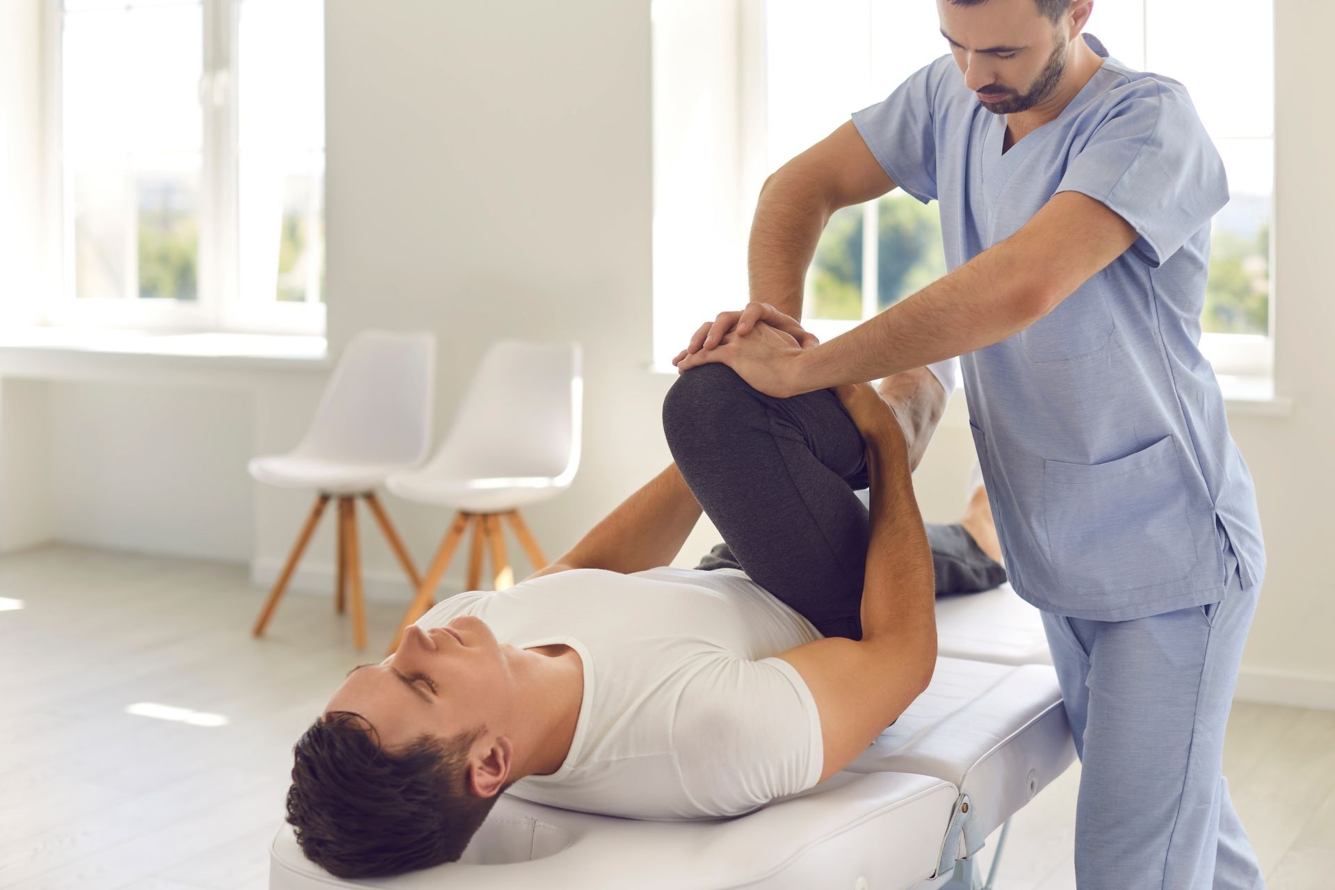
Treatment options for managing patellofemoral instability may include conservative measures such as physical therapy to strengthen the surrounding muscles, bracing or taping to provide additional support, and activity modification to avoid aggravating movements. In cases where conservative treatments are ineffective, surgical interventions like realignment procedures or ligament reconstructions may be necessary to stabilize the patella and prevent further dislocations.

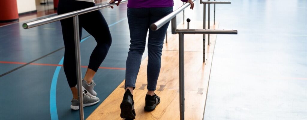
Physical therapy plays a crucial role in the treatment of patellofemoral instability by focusing on strengthening the quadriceps, hamstrings, and hip muscles to improve joint stability and alignment. Therapists may also incorporate exercises to improve flexibility, balance, and proprioception to enhance overall knee function and reduce the risk of future dislocations. Consistent participation in a structured physical therapy program can help individuals regain strength and function in the affected knee.
Specific exercises and stretches that can help prevent patellofemoral instability from worsening include quad sets, straight leg raises, clamshells, and hip abduction exercises to target the muscles that support the patella. Stretching exercises for the quadriceps, hamstrings, and iliotibial band can also help improve flexibility and reduce tension around the knee joint. It is essential to perform these exercises under the guidance of a healthcare provider or physical therapist to ensure proper form and progression to prevent exacerbating instability.
Orthopedic physical therapy has the potential to positively impact bone density in postmenopausal women by promoting weight-bearing exercises, resistance training, and balance exercises. These interventions can help stimulate bone formation, increase bone mineral density, and reduce the risk of osteoporosis-related fractures. By targeting specific muscle groups and joints through tailored exercise programs, physical therapists can improve overall bone health and strength in postmenopausal women. Additionally, the incorporation of weight-bearing activities can enhance bone remodeling processes, leading to improved bone density over time. Regular monitoring and adjustments to the physical therapy regimen can further optimize outcomes and support long-term bone health in this population.
Orthopedic physical therapy approaches the rehabilitation of individuals with complex regional pain syndrome (CRPS) by focusing on a multidisciplinary treatment plan that addresses the specific needs of the patient. This approach may include a combination of manual therapy techniques, such as joint mobilization and soft tissue mobilization, to improve range of motion and reduce pain. Additionally, therapeutic exercises targeting muscle imbalances and proprioception deficits are utilized to improve functional movement patterns. Modalities such as heat, ice, and electrical stimulation may also be incorporated to help manage pain and inflammation. Education on pain management strategies, activity modification, and ergonomic principles is provided to empower the individual in self-management of their condition. Overall, orthopedic physical therapy aims to improve the individual's quality of life and functional abilities while addressing the complexities of CRPS.
Orthopedic physical therapy for individuals with pes planus focuses on addressing muscle imbalances through targeted exercises and interventions. This may include strengthening the intrinsic foot muscles, such as the flexor hallucis brevis and abductor hallucis, to improve arch support and stability. Additionally, exercises to strengthen the posterior tibialis muscle can help correct overpronation and improve foot alignment. Stretching exercises for tight calf muscles and the plantar fascia can also be incorporated to address any contributing factors to the muscle imbalances. Orthopedic physical therapists may also utilize manual therapy techniques to release tight muscles and improve joint mobility in the foot and ankle complex. By addressing these muscle imbalances, individuals with pes planus can improve their overall foot function and reduce the risk of pain and injury.
Common exercises for rehabilitating a torn ACL in orthopedic physical therapy typically include a combination of strengthening, flexibility, and balance exercises. Some specific exercises may include leg presses, hamstring curls, calf raises, and squats to strengthen the muscles around the knee. Flexibility exercises such as hamstring stretches and quad stretches can help improve range of motion. Balance exercises like single-leg stands and stability ball exercises can help improve proprioception and reduce the risk of future injury. Additionally, functional exercises such as lunges and step-ups may be incorporated to help patients regain strength and stability for everyday activities. It is important for physical therapists to tailor the exercise program to each individual's specific needs and stage of recovery to ensure optimal outcomes.
Orthopedic physical therapy for pediatric patients with congenital musculoskeletal disorders focuses on addressing the specific needs of this population through a combination of specialized techniques and interventions. These may include exercises to improve strength, flexibility, and range of motion, as well as manual therapy to address joint stiffness and muscle imbalances. Additionally, orthopedic physical therapists may utilize modalities such as electrical stimulation or ultrasound to help manage pain and promote healing. Education plays a crucial role in treatment, with therapists providing guidance on proper body mechanics, adaptive equipment, and home exercise programs. By tailoring treatment plans to the unique needs of pediatric patients with congenital musculoskeletal disorders, orthopedic physical therapy aims to improve functional outcomes and enhance quality of life for these individuals.
In orthopedic physical therapy, addressing trigger points typically involves a combination of manual therapy techniques such as myofascial release, trigger point release, and deep tissue massage. Therapists may also utilize modalities like ultrasound, electrical stimulation, or dry needling to help alleviate muscle tension and pain associated with trigger points. Additionally, therapeutic exercises focusing on stretching, strengthening, and neuromuscular re-education can help prevent trigger points from recurring. Education on proper posture, ergonomics, and self-care strategies may also be provided to empower patients in managing their trigger points outside of therapy sessions. Overall, a comprehensive approach tailored to the individual's specific needs and goals is essential in effectively addressing trigger points in orthopedic physical therapy.
Orthopedic physical therapy for individuals with Duchenne muscular dystrophy focuses on addressing muscle weakness through a combination of targeted exercises, stretching techniques, and functional training. These interventions aim to improve muscle strength, flexibility, and overall function in order to enhance mobility and quality of life for patients with this progressive neuromuscular disorder. By incorporating resistance training, proprioceptive exercises, and neuromuscular re-education, physical therapists can help individuals with Duchenne muscular dystrophy maintain muscle mass, prevent contractures, and optimize movement patterns. Additionally, orthopedic physical therapy may involve the use of assistive devices, orthotics, and adaptive equipment to support weakened muscles and facilitate safe and efficient movement. Overall, a comprehensive approach to addressing muscle weakness in individuals with Duchenne muscular dystrophy can help to slow disease progression and improve functional outcomes.
Exercises that are recommended for improving hip internal rotation strength include clamshells, hip internal rotation with resistance bands, seated hip internal rotation, hip internal rotation with a stability ball, and hip internal rotation with a foam roller. These exercises target the muscles responsible for hip internal rotation, such as the gluteus medius, gluteus minimus, and tensor fasciae latae. Strengthening these muscles can help improve hip stability, reduce the risk of injury, and enhance overall lower body strength and function. It is important to perform these exercises with proper form and gradually increase resistance to continue challenging the muscles and promoting strength gains in hip internal rotation.