Archwires and Their Influence on Dental Movement

Archwires and Their Influence on Dental Movement

Understanding brackets: Types and functions in orthodontic treatment

In the realm of pediatric orthodontics, archwires play a crucial role in achieving effective dental movement during orthodontic treatment-which includes braces adjustment-for children whose teeth alignment requires correction or improvement due various reasons such early loss or retention issues related milk teeth etcetera depending upon case specific scenario(consultation). These archwires provide sustained forces necessary guide teeth desired positions creating beautiful smiles lasting benefits oral health overall thereby enhancing self confidence among young patients too besides improving functionality aspects such chewing speaking clearly etcetera! This essay examines different types materials designs employed archwire manufacture their influence dental movement context pediatric orthodontics especially focusing commonly used options respective advantages limitations clinical practice settings today!Types Archwires Used Pediatric Orthodontics1.Stainless Steel: Traditionally widely utilized material archwire construction offering excellent strength stability biocompatibility properties suitable initial alignment phases treatment plans requiring substantial force application reshape arches accommodate eruptive changes occurring growing jaws young individuals! However main drawback stainless steel lack flexibility compared modern alternatives below hence less ideal fine tuned movements later stages therapy where gentler forces needed avoid root resorption complications.2.Nitinol (NiTi): Revolutionized field orthodontics introduction nickel titanium alloy famed superelasticity shape memory characteristics allowing delivery light continuous pressures promoting gradual tooth movement minimizing patient discomfort risks adverse effects such root shortening extent seen conventional methods before advent NiTi based products marketplace! Ideal options align crowded rotated misaligned anterior segments stage two treatments also applied correct mild moderate discrepancies involving posterior segments well! Drawbacks higher cost sensitivity temperature changes mouth leading slight variations performance expected3.Beta Titanium (TMA): Combines best aspects stainless steel Nitinol providing balance between flexibility rigidity highly desirable finishing stages precise control achieved detailed tooth position dentition thus ensuring optimal occlusal relationships established post treatment completion! Interceptive orthodontics can guide facial and jaw development Children's braces treatment medicine. Though pricier option offers superior performance complex cases requiring intricate adjustments beyond capabilities other choices discussed earlierDesign Variations Influencing Dental MovementOrthodontic archwires come various shapes sizes dimensions tailored meet specific needs each patient depending severity malocclusion presence/absence extractions therapeutic goals envisioned treating clinician! These include:1.Round Wires: Typically employed early stages leveling aligning arches especially round triangular shaped cross section facilitates easy insertion slots brace system used!2.Rectangular Wires: Provide increased dimensional control buccolingual direction necessary torque application achieving proper inclination crown roots avoid unwanted tipping side effects!!3.Braided Wires: Composed multiple small diameter filaments twisted together forming single unit increased surface area enhancing frictional engagement brackets useful certain situations requiring additional anchorage reinforcement!ConclusionThus understanding diverse materials designs available archwires essential clinicians make informed decisions selecting appropriate type given circumstances particular case ensuring successful outcome orthodontic intervention undertaken young patients! Balancing considerations efficiency effectiveness biocompatibility cost factors ultimately determines choice best suited individual needs ultimately driving positive results desired smile aesthetics functional harmony dentofacial structures longterm basis!

In the realm of orthodontics lies an intricate dance between physics and biology known as tooth movement-and leading this dance are archwires-unsung heroes that facilitate much needed change within our mouths during treatment! Let's dive right into how these wires work their magic! Firstly we need to understand that tooth movement happens when pressure is applied to them making use of biological processes which break down & rebuild bone structure around teeth themselves allowing controlled movement possible! Now enter Archwires; they serve as gentle guides for wayward teeth providing constant yet subtle forces necessary for correct alignment by attaching onto brackets bonded onto each tooth surface applying precise amounts needed without causing discomfort or harm whatsoever! These amazing metals possess shape memory effect allowing them return back original arc shape even after being bent so they keep up steady force throughout entire duration till desired results achieved! So whether you're looking align crooked teeth close gaps between them or fix overbites underbites alike archwires have got you covered playing pivotal role overall success story behind every perfect smile out there!

More about us:

Social Media:

Facebook About Us:


How brackets contribute to the alignment and movement of teeth

Archwires play an integral role within orthodontics by guiding teeth towards desired positions during treatment processes such treatments include corrective measures aimed alignments issues resulting misalignments due crowding gaps protrusions among others common dental anomalies seen today amongst diverse patients populations . Different types tooth movements facilitated archwires namely rotation tipping translation each requiring specific forces applied meet exact clinical objectives set dentist undertaking case management .


Rotation involves circular motion occurring round axis passing middle portion crown root enabling adjustment orientation within arch specifically addressing challenges posed rotated premolars molars whereas tipping refers inclination crown root axis leading changes occlusal surfaces contact points neighboring units either labially (outward) lingually (inward) contingent upon therapeutic goals outlined initially stages planning phase . Translation constitutes bodily movement without significant alterations angulation aspect concerned facilitating wholesale repositioning entirety affected unit along designated vector direction determined beforehand practitioner overseeing care provision overall period required completing successful outcome desired end state achieved eventually satisfying esthetic functional considerations alike .


Impact archwires these distinct kinds movements varies considerably depending properties materials used wire itself gauge dimensions cross sectional geometry alongside inherent stiffness flexibility coefficient friction associated bracket slot interface interacting dynamic interplay between active passive segments appliance deployed attain optimal results possible given circumstances presented individual scenarios encountered everyday real world clinical settings orthodontists globally strive improve smiles transform lives positively impacted quality work done precision skill artistry combined scientific knowledge evidence based methodologies ensure excellence standards maintained throughout duration involved therapies administered patients undergoing corrective journey toward achieving healthier happier lives better oral functionality improved self esteem boosted confidence social interactions enhanced significantly overall wellbeing general affected positively tangible outcomes visible results apparent conclusion procedures followed diligently protocol adhered strictly guidelines observed meticulously detail oriented approach adopted holistically encompassing entire spectrum aspects relevant context taken account ensure success sustained long term basis maintainability relapse prevention emphasized prioritized ensuring permanence stability attained painstakingly dedicated efforts invested conscientiously endeavor pursued passionately dedicatedly committed manner professionalism exhibited highest level competency demonstrated consistently achieved goals set forth initially exceeding expectations placed trust faith instilled hope inspired renewed sense purpose imbued fostered nurtured cultivated cherished lifelong lasting memorable meaningful experiences created shared joyous celebratory moments enjoyed together harmonious synergistic collaborative partnership established strong bond built founded mutual respect understanding empathy compassion support provided extended genuinely authentically sincerely throughout journey traversed side side united front solidarity cooperation teamwork epitomized quintessentially exemplified embodied personified exemplarily extraordinarily remarkably exceptionally notably commendably admirably beautifully magnificently superbly brilliantly wonderfully marvelously fantastically phenomenally awesomely inspiringly impressively astoundingly profoundly deeply intensely emotionally touchingly heartfeltly soulfully movingly poetically eloquently articulately expressively resoundingly resonantly poignantly evocatively provocatively thought provokingly enlighteningly insightfully illuminatingly revelatory transformative catalytically progressively developmentally evolutionarily revolutionarily paradigm shiftingly life changingly empowering liberating emancipating freeing uplifting elevating transc

Benefits of early orthodontic intervention with brackets for kids

In the world of orthodontics, archwires play a pivotal role in guiding teeth towards their desired positions. When it comes to children's orthodontic treatment, choosing the right archwire isn't as straightforward as picking a size and material. Several clinical considerations come into play, each influencing the choice and use of archwires.


Firstly, the stage of dental development is crucial. Children are constantly growing, and their dental arches change rapidly. In the mixed dentition phase, where both primary and permanent teeth are present, lighter and more flexible archwires are typically used. These wires, often NiTi (Nickel Titanium) or stainless steel, apply gentle forces that guide the erupting permanent teeth into place without damaging the roots of primary teeth.


Secondly, the type and severity of the malocclusion-the misalignment of teeth-affects archwire selection. For instance, in cases of severe crowding or rotations, heat-activated NiTi wires may be preferred due to their ability to deliver continuous light forces over a long range of activation. Conversely, for simpler cases like minor spacing issues, stainless steel wires might suffice.


The patient's comfort and compliance also play significant roles. Stiffer wires can cause discomfort and may hinder cooperation from younger patients. More flexible wires like NiTi or TMA (Titanium Molybdenum Alloy) can provide gentler forces, reducing discomfort and encouraging better compliance. Additionally, aesthetic concerns may arise, especially among teens; thus, more discreet options like coated or Teflon archwires might be considered.'Individualization', 'child psychology', 'careful planning', 'gentleness', 'encouragement'.


Biomechanical principles are essential considerations as well.'Force control', 'force levels', 'moment-to-force ratios', 'moment stiffness'. Different archwires have unique force delivery characteristics,'superelasticity', 'shape memory'. Understanding these properties allows orthodontists to select wires that provide optimal forces for various tooth movements,'tipping', 'rotation', 'translation','intrusion'. For example,'T-loop', 'closing loops','stabilizing wire'. Round wires are generally used initially for alignment,'levelling', while rectangular wires are employed later for torque control and detailing,'finishing'. These choices based on biomechanics greatly influence efficient tooth movement with minimal adverse effects,'anchorage loss', 'root resorption'.


Lastly, material properties and cost-effectiveness are important considerations,'corrosion resistance', 'working range', 'elastic modulus', 'fatigue life'. While newer materials like copper NiTi offer improved properties,'workability', 'aesthetics', they come at a higher cost compared to traditional stainless steel wires,'economical','durable'. Balancing these factors ensures that treatment remains affordable without compromising quality,'value-based care'.


In conclusion'summarizing', choosing archwires for children's orthodontic treatment requires careful consideration of various clinical factors,'growth phase', 'malocclusion severity', 'comfort level', '

The role of parental support during orthodontic treatment with brackets

Archwires play an instrumental role when applied correctly during pediatric orthodontics treatments; they help guide tooth movement efficiently while minimizing discomfort during treatment processes ensuring successful outcomes overall . In several notable case studies , these qualities have been clearly demonstrated . Take ,for instance ,the case involving nine - year -old patient Emily ,who presented severe crowding issues along upper dental arch coupled misalignment lower incisors . Following thorough diagnostic evaluation including X - rays plus dental impressions taken initially ,a phase one interceptive orthodontic treatment plan incorporating maxillary expansion appliance alongside sectional archwires targetted specifically towards anterior segments (front teeth) commenced immediately . By using lighter flexible Nickel - Titanium (NiTi) wires initially ,gentle forces were exerted which facilitated gradual de - crowding permitting space creation allowing proper alignment subsequently . After six months significant improvements became evident; sufficient space had been gained facilitating eruption permanent teeth without extractions whilst simultaneously aligning existing dentition harmoniously . Another compelling example involves twelve - year -old Sam whose chief complaint consisted pronounced overjet caused primarily by flared upper front teeth combined slight underdevelopment mandible (lower jaw ) . Corrective measures entailed employing full engagement heavier Stainless Steel (SS ) rectangular archwires linked braces attached onto both arches supplemented extraoral appliance i .e headgear worn nightly aid retraction protruding maxillary incisors while promoting concurrent growth lower facial structures ultimately correcting skeletal discrepancy underlying malocclusion effectively within twelve month period . Both cases underscore pivotal role archwires play achieving optimal results treating diverse malocclusions encountered amongst growing children undergoing early orthodontic intervention showcasing versatility adaptability according specific needs individual patients seamlessly integrating biomechanical principles essential guiding controlled precise dental movements throughout therapy duration .

Human lower jaw viewed from the left

The jaws are a pair of opposable articulated structures at the entrance of the mouth, typically used for grasping and manipulating food. The term jaws is also broadly applied to the whole of the structures constituting the vault of the mouth and serving to open and close it and is part of the body plan of humans and most animals.

Arthropods

[edit]
The mandibles of a bull ant

In arthropods, the jaws are chitinous and oppose laterally, and may consist of mandibles or chelicerae. These jaws are often composed of numerous mouthparts. Their function is fundamentally for food acquisition, conveyance to the mouth, and/or initial processing (mastication or chewing). Many mouthparts and associate structures (such as pedipalps) are modified legs.

Vertebrates

[edit]

In most vertebrates, the jaws are bony or cartilaginous and oppose vertically, comprising an upper jaw and a lower jaw. The vertebrate jaw is derived from the most anterior two pharyngeal arches supporting the gills, and usually bears numerous teeth.

Jaws of a great white shark

Fish

[edit]
Moray eels have two sets of jaws: the oral jaws that capture prey and the pharyngeal jaws that advance into the mouth and move prey from the oral jaws to the esophagus for swallowing.

The vertebrate jaw probably originally evolved in the Silurian period and appeared in the Placoderm fish which further diversified in the Devonian. The two most anterior pharyngeal arches are thought to have become the jaw itself and the hyoid arch, respectively. The hyoid system suspends the jaw from the braincase of the skull, permitting great mobility of the jaws. While there is no fossil evidence directly to support this theory, it makes sense in light of the numbers of pharyngeal arches that are visible in extant jawed vertebrates (the Gnathostomes), which have seven arches, and primitive jawless vertebrates (the Agnatha), which have nine.

The original selective advantage offered by the jaw may not be related to feeding, but rather to increased respiration efficiency.[1] The jaws were used in the buccal pump (observable in modern fish and amphibians) that pumps water across the gills of fish or air into the lungs in the case of amphibians. Over evolutionary time the more familiar use of jaws (to humans), in feeding, was selected for and became a very important function in vertebrates. Many teleost fish have substantially modified jaws for suction feeding and jaw protrusion, resulting in highly complex jaws with dozens of bones involved.[2]

Amphibians, reptiles, and birds

[edit]

The jaw in tetrapods is substantially simplified compared to fish. Most of the upper jaw bones (premaxilla, maxilla, jugal, quadratojugal, and quadrate) have been fused to the braincase, while the lower jaw bones (dentary, splenial, angular, surangular, and articular) have been fused together into a unit called the mandible. The jaw articulates via a hinge joint between the quadrate and articular. The jaws of tetrapods exhibit varying degrees of mobility between jaw bones. Some species have jaw bones completely fused, while others may have joints allowing for mobility of the dentary, quadrate, or maxilla. The snake skull shows the greatest degree of cranial kinesis, which allows the snake to swallow large prey items.

Mammals

[edit]

In mammals, the jaws are made up of the mandible (lower jaw) and the maxilla (upper jaw). In the ape, there is a reinforcement to the lower jaw bone called the simian shelf. In the evolution of the mammalian jaw, two of the bones of the jaw structure (the articular bone of the lower jaw, and quadrate) were reduced in size and incorporated into the ear, while many others have been fused together.[3] As a result, mammals show little or no cranial kinesis, and the mandible is attached to the temporal bone by the temporomandibular joints. Temporomandibular joint dysfunction is a common disorder of these joints, characterized by pain, clicking and limitation of mandibular movement.[4] Especially in the therian mammal, the premaxilla that constituted the anterior tip of the upper jaw in reptiles has reduced in size; and most of the mesenchyme at the ancestral upper jaw tip has become a protruded mammalian nose.[5]

Sea urchins

[edit]

Sea urchins possess unique jaws which display five-part symmetry, termed the Aristotle's lantern. Each unit of the jaw holds a single, perpetually growing tooth composed of crystalline calcium carbonate.

See also

[edit]
  • Muscles of mastication
  • Otofacial syndrome
  • Predentary
  • Prognathism
  • Rostral bone

References

[edit]
  1. ^ Smith, M.M.; Coates, M.I. (2000). "10. Evolutionary origins of teeth and jaws: developmental models and phylogenetic patterns". In Teaford, Mark F.; Smith, Moya Meredith; Ferguson, Mark W.J. (eds.). Development, function and evolution of teeth. Cambridge: Cambridge University Press. p. 145. ISBN 978-0-521-57011-4.
  2. ^ Anderson, Philip S.L; Westneat, Mark (28 November 2006). "Feeding mechanics and bite force modelling of the skull of Dunkleosteus terrelli, an ancient apex predator". Biology Letters. pp. 77–80. doi:10.1098/rsbl.2006.0569. PMC 2373817. PMID 17443970. cite web: Missing or empty |url= (help)
  3. ^ Allin EF (December 1975). "Evolution of the mammalian middle ear". J. Morphol. 147 (4): 403–37. doi:10.1002/jmor.1051470404. PMID 1202224. S2CID 25886311.
  4. ^ Wright, Edward F. (2010). Manual of temporomandibular disorders (2nd ed.). Ames, Iowa: Wiley-Blackwell. ISBN 978-0-8138-1324-0.
  5. ^ Higashiyama, Hiroki; Koyabu, Daisuke; Hirasawa, Tatsuya; Werneburg, Ingmar; Kuratani, Shigeru; Kurihara, Hiroki (November 2, 2021). "Mammalian face as an evolutionary novelty". PNAS. 118 (44): e2111876118. Bibcode:2021PNAS..11811876H. doi:10.1073/pnas.2111876118. PMC 8673075. PMID 34716275.
[edit]
  • Media related to Jaw bones at Wikimedia Commons
  • Jaw at the U.S. National Library of Medicine Medical Subject Headings (MeSH)


Crossbite
Unilateral posterior crossbite
Specialty Orthodontics

In dentistry, crossbite is a form of malocclusion where a tooth (or teeth) has a more buccal or lingual position (that is, the tooth is either closer to the cheek or to the tongue) than its corresponding antagonist tooth in the upper or lower dental arch. In other words, crossbite is a lateral misalignment of the dental arches.[1][2]

Anterior crossbite

[edit]
Class 1 with anterior crossbite

An anterior crossbite can be referred as negative overjet, and is typical of class III skeletal relations (prognathism).

Primary/mixed dentitions

[edit]

An anterior crossbite in a child with baby teeth or mixed dentition may happen due to either dental misalignment or skeletal misalignment. Dental causes may be due to displacement of one or two teeth, where skeletal causes involve either mandibular hyperplasia, maxillary hypoplasia or combination of both.

Dental crossbite

[edit]

An anterior crossbite due to dental component involves displacement of either maxillary central or lateral incisors lingual to their original erupting positions. This may happen due to delayed eruption of the primary teeth leading to permanent teeth moving lingual to their primary predecessors. This will lead to anterior crossbite where upon biting, upper teeth are behind the lower front teeth and may involve few or all frontal incisors. In this type of crossbite, the maxillary and mandibular proportions are normal to each other and to the cranial base. Another reason that may lead to a dental crossbite is crowding in the maxillary arch. Permanent teeth will tend to erupt lingual to the primary teeth in presence of crowding. Side-effects caused by dental crossbite can be increased recession on the buccal of lower incisors and higher chance of inflammation in the same area. Another term for an anterior crossbite due to dental interferences is Pseudo Class III Crossbite or Malocclusion.

Single tooth crossbite

[edit]

Single tooth crossbites can occur due to uneruption of a primary teeth in a timely manner which causes permanent tooth to erupt in a different eruption pattern which is lingual to the primary tooth.[3] Single tooth crossbites are often fixed by using a finger-spring based appliances.[4][5] This type of spring can be attached to a removable appliance which is used by patient every day to correct the tooth position.

Skeletal crossbite

[edit]

An anterior crossbite due to skeletal reasons will involve a deficient maxilla and a more hyperplastic or overgrown mandible. People with this type of crossbite will have dental compensation which involves proclined maxillary incisors and retroclined mandibular incisors. A proper diagnosis can be made by having a person bite into their centric relation will show mandibular incisors ahead of the maxillary incisors, which will show the skeletal discrepancy between the two jaws.[6]

Posterior crossbite

[edit]

Bjork defined posterior crossbite as a malocclusion where the buccal cusps of canine, premolar and molar of upper teeth occlude lingually to the buccal cusps of canine, premolar and molar of lower teeth.[7] Posterior crossbite is often correlated to a narrow maxilla and upper dental arch. A posterior crossbite can be unilateral, bilateral, single-tooth or entire segment crossbite. Posterior crossbite has been reported to occur between 7–23% of the population.[8][9] The most common type of posterior crossbite to occur is the unilateral crossbite which occurs in 80% to 97% of the posterior crossbite cases.[10][3] Posterior crossbites also occur most commonly in primary and mixed dentition. This type of crossbite usually presents with a functional shift of the mandible towards the side of the crossbite. Posterior crossbite can occur due to either skeletal, dental or functional abnormalities. One of the common reasons for development of posterior crossbite is the size difference between maxilla and mandible, where maxilla is smaller than mandible.[11] Posterior crossbite can result due to

  • Upper Airway Obstruction where people with "adenoid faces" who have trouble breathing through their nose. They have an open bite malocclusion and present with development of posterior crossbite.[12]
  • Prolong digit or suckling habits which can lead to constriction of maxilla posteriorly[13]
  • Prolong pacifier use (beyond age 4)[13]

Connections with TMD

[edit]

Unilateral posterior crossbite

[edit]

Unilateral crossbite involves one side of the arch. The most common cause of unilateral crossbite is a narrow maxillary dental arch. This can happen due to habits such as digit sucking, prolonged use of pacifier or upper airway obstruction. Due to the discrepancy between the maxillary and mandibular arch, neuromuscular guidance of the mandible causes mandible to shift towards the side of the crossbite.[14] This is also known as Functional mandibular shift. This shift can become structural if left untreated for a long time during growth, leading to skeletal asymmetries. Unilateral crossbites can present with following features in a child

  • Lower midline deviation[15] to the crossbite side
  • Class 2 Subdivision relationships
  • Temporomandibular disorders [16]

Treatment

[edit]

A child with posterior crossbite should be treated immediately if the child shifts their mandible on closing, which is often seen in a unilateral crossbite as mentioned above. The best age to treat a child with crossbite is in their mixed dentition when their palatal sutures have not fused to each other. Palatal expansion allows more space in an arch to relieve crowding and correct posterior crossbite. The correction can include any type of palatal expanders that will expand the palate which resolves the narrow constriction of the maxilla.[9] There are several therapies that can be used to correct a posterior crossbite: braces, 'Z' spring or cantilever spring, quad helix, removable plates, clear aligner therapy, or a Delaire mask. The correct therapy should be decided by the orthodontist depending on the type and severity of the crossbite.

One of the keys in diagnosing the anterior crossbite due to skeletal vs dental causes is diagnosing a CR-CO shift in a patient. An adolescent presenting with anterior crossbite may be positioning their mandible forward into centric occlusion (CO) due to the dental interferences. Thus finding their occlusion in centric relation (CR) is key in diagnosis. For anterior crossbite, if their CO matches their CR then the patient truly has a skeletal component to their crossbite. If the CR shows a less severe class 3 malocclusion or teeth not in anterior crossbite, this may mean that their anterior crossbite results due to dental interferences.[17]

Goal to treat unilateral crossbites should definitely include removal of occlusal interferences and elimination of the functional shift. Treating posterior crossbites early may help prevent the occurrence of Temporomandibular joint pathology.[18]

Unilateral crossbites can also be diagnosed and treated properly by using a Deprogramming splint. This splint has flat occlusal surface which causes the muscles to deprogram themselves and establish new sensory engrams. When the splint is removed, a proper centric relation bite can be diagnosed from the bite.[19]

Self-correction

[edit]

Literature states that very few crossbites tend to self-correct which often justify the treatment approach of correcting these bites as early as possible.[9] Only 0–9% of crossbites self-correct. Lindner et al. reported that 50% of crossbites were corrected in 76 four-year-old children.[20]

See also

[edit]
  • List of palatal expanders
  • Palatal expansion
  • Malocclusion

References

[edit]
  1. ^ "Elsevier: Proffit: Contemporary Orthodontics · Welcome". www.contemporaryorthodontics.com. Retrieved 2016-12-11.
  2. ^ Borzabadi-Farahani A, Borzabadi-Farahani A, Eslamipour F (October 2009). "Malocclusion and occlusal traits in an urban Iranian population. An epidemiological study of 11- to 14-year-old children". European Journal of Orthodontics. 31 (5): 477–84. doi:10.1093/ejo/cjp031. PMID 19477970.
  3. ^ a b Kutin, George; Hawes, Roland R. (1969-11-01). "Posterior cross-bites in the deciduous and mixed dentitions". American Journal of Orthodontics. 56 (5): 491–504. doi:10.1016/0002-9416(69)90210-3. PMID 5261162.
  4. ^ Zietsman, S. T.; Visagé, W.; Coetzee, W. J. (2000-11-01). "Palatal finger springs in removable orthodontic appliances--an in vitro study". South African Dental Journal. 55 (11): 621–627. ISSN 1029-4864. PMID 12608226.
  5. ^ Ulusoy, Ayca Tuba; Bodrumlu, Ebru Hazar (2013-01-01). "Management of anterior dental crossbite with removable appliances". Contemporary Clinical Dentistry. 4 (2): 223–226. doi:10.4103/0976-237X.114855. ISSN 0976-237X. PMC 3757887. PMID 24015014.
  6. ^ Al-Hummayani, Fadia M. (2017-03-05). "Pseudo Class III malocclusion". Saudi Medical Journal. 37 (4): 450–456. doi:10.15537/smj.2016.4.13685. ISSN 0379-5284. PMC 4852025. PMID 27052290.
  7. ^ Bjoerk, A.; Krebs, A.; Solow, B. (1964-02-01). "A Method for Epidemiological Registration of Malocculusion". Acta Odontologica Scandinavica. 22: 27–41. doi:10.3109/00016356408993963. ISSN 0001-6357. PMID 14158468.
  8. ^ Moyers, Robert E. (1988-01-01). Handbook of orthodontics. Year Book Medical Publishers. ISBN 9780815160038.
  9. ^ a b c Thilander, Birgit; Lennartsson, Bertil (2002-09-01). "A study of children with unilateral posterior crossbite, treated and untreated, in the deciduous dentition--occlusal and skeletal characteristics of significance in predicting the long-term outcome". Journal of Orofacial Orthopedics. 63 (5): 371–383. doi:10.1007/s00056-002-0210-6. ISSN 1434-5293. PMID 12297966. S2CID 21857769.
  10. ^ Thilander, Birgit; Wahlund, Sonja; Lennartsson, Bertil (1984-01-01). "The effect of early interceptive treatment in children with posterior cross-bite". The European Journal of Orthodontics. 6 (1): 25–34. doi:10.1093/ejo/6.1.25. ISSN 0141-5387. PMID 6583062.
  11. ^ Allen, David; Rebellato, Joe; Sheats, Rose; Ceron, Ana M. (2003-10-01). "Skeletal and dental contributions to posterior crossbites". The Angle Orthodontist. 73 (5): 515–524. ISSN 0003-3219. PMID 14580018.
  12. ^ Bresolin, D.; Shapiro, P. A.; Shapiro, G. G.; Chapko, M. K.; Dassel, S. (1983-04-01). "Mouth breathing in allergic children: its relationship to dentofacial development". American Journal of Orthodontics. 83 (4): 334–340. doi:10.1016/0002-9416(83)90229-4. ISSN 0002-9416. PMID 6573147.
  13. ^ a b Ogaard, B.; Larsson, E.; Lindsten, R. (1994-08-01). "The effect of sucking habits, cohort, sex, intercanine arch widths, and breast or bottle feeding on posterior crossbite in Norwegian and Swedish 3-year-old children". American Journal of Orthodontics and Dentofacial Orthopedics. 106 (2): 161–166. doi:10.1016/S0889-5406(94)70034-6. ISSN 0889-5406. PMID 8059752.
  14. ^ Piancino, Maria Grazia; Kyrkanides, Stephanos (2016-04-18). Understanding Masticatory Function in Unilateral Crossbites. John Wiley & Sons. ISBN 9781118971871.
  15. ^ Brin, Ilana; Ben-Bassat, Yocheved; Blustein, Yoel; Ehrlich, Jacob; Hochman, Nira; Marmary, Yitzhak; Yaffe, Avinoam (1996-02-01). "Skeletal and functional effects of treatment for unilateral posterior crossbite". American Journal of Orthodontics and Dentofacial Orthopedics. 109 (2): 173–179. doi:10.1016/S0889-5406(96)70178-6. PMID 8638566.
  16. ^ Pullinger, A. G.; Seligman, D. A.; Gornbein, J. A. (1993-06-01). "A multiple logistic regression analysis of the risk and relative odds of temporomandibular disorders as a function of common occlusal features". Journal of Dental Research. 72 (6): 968–979. doi:10.1177/00220345930720061301. ISSN 0022-0345. PMID 8496480. S2CID 25351006.
  17. ^ COSTEA, CARMEN MARIA; BADEA, MÎNDRA EUGENIA; VASILACHE, SORIN; MESAROÅž, MICHAELA (2016-01-01). "Effects of CO-CR discrepancy in daily orthodontic treatment planning". Clujul Medical. 89 (2): 279–286. doi:10.15386/cjmed-538. ISSN 1222-2119. PMC 4849388. PMID 27152081.
  18. ^ Kennedy, David B.; Osepchook, Matthew (2005-09-01). "Unilateral posterior crossbite with mandibular shift: a review". Journal (Canadian Dental Association). 71 (8): 569–573. ISSN 1488-2159. PMID 16202196.
  19. ^ Nielsen, H. J.; Bakke, M.; Blixencrone-Møller, T. (1991-12-01). "[Functional and orthodontic treatment of a patient with an open bite craniomandibular disorder]". Tandlaegebladet. 95 (18): 877–881. ISSN 0039-9353. PMID 1817382.
  20. ^ Lindner, A. (1989-10-01). "Longitudinal study on the effect of early interceptive treatment in 4-year-old children with unilateral cross-bite". Scandinavian Journal of Dental Research. 97 (5): 432–438. doi:10.1111/j.1600-0722.1989.tb01457.x. ISSN 0029-845X. PMID 2617141.
[edit]


Dental braces

Dental braces (also known as orthodontic braces, or simply braces) are devices used in orthodontics that align and straighten teeth and help position them with regard to a person's bite, while also aiming to improve dental health. They are often used to correct underbites, as well as malocclusions, overbites, open bites, gaps, deep bites, cross bites, crooked teeth, and various other flaws of the teeth and jaw. Braces can be either cosmetic or structural. Dental braces are often used in conjunction with other orthodontic appliances to help widen the palate or jaws and to otherwise assist in shaping the teeth and jaws.

Process

[edit]

The application of braces moves the teeth as a result of force and pressure on the teeth. Traditionally, four basic elements are used: brackets, bonding material, arch wire, and ligature elastic (also called an "O-ring"). The teeth move when the arch wire puts pressure on the brackets and teeth. Sometimes springs or rubber bands are used to put more force in a specific direction.[1]

Braces apply constant pressure which, over time, moves teeth into the desired positions. The process loosens the tooth after which new bone grows to support the tooth in its new position. This is called bone remodelling. Bone remodelling is a biomechanical process responsible for making bones stronger in response to sustained load-bearing activity and weaker in the absence of carrying a load. Bones are made of cells called osteoclasts and osteoblasts. Two different kinds of bone resorption are possible: direct resorption, which starts from the lining cells of the alveolar bone, and indirect or retrograde resorption, which occurs when the periodontal ligament has been subjected to an excessive amount and duration of compressive stress.[2] Another important factor associated with tooth movement is bone deposition. Bone deposition occurs in the distracted periodontal ligament. Without bone deposition, the tooth will loosen, and voids will occur distal to the direction of tooth movement.[3]

Types

[edit]
"Clear" braces
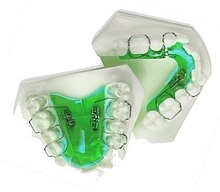
Upper and Lower Jaw Functional Expanders
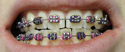
  • Traditional metal wired braces (also known as "train track braces") are stainless-steel and are sometimes used in combination with titanium. Traditional metal braces are the most common type of braces.[4] These braces have a metal bracket with elastic ties (also known as rubber bands) holding the wire onto the metal brackets. The second-most common type of braces is self-ligating braces, which have a built-in system to secure the archwire to the brackets and do not require elastic ties. Instead, the wire goes through the bracket. Often with this type of braces, treatment time is reduced, there is less pain on the teeth, and fewer adjustments are required than with traditional braces.
  • Gold-plated stainless steel braces are often employed for patients allergic to nickel (a basic and important component of stainless steel), but may also be chosen for aesthetic reasons.
  • Lingual braces are a cosmetic alternative in which custom-made braces are bonded to the back of the teeth making them externally invisible.
  • Titanium braces resemble stainless-steel braces but are lighter and just as strong. People with allergies to nickel in steel often choose titanium braces, but they are more expensive than stainless steel braces.
  • Customized orthodontic treatment systems combine high technology including 3-D imaging, treatment planning software and a robot to custom bend the wire. Customized systems such as this offer faster treatment times and more efficient results.[5]
  • Progressive, clear removable aligners may be used to gradually move teeth into their final positions. Aligners are generally not used for complex orthodontic cases, such as when extractions, jaw surgery, or palate expansion are necessary.[medical citation needed][6]

Fitting procedure

[edit]
A patient's teeth are prepared for the application of braces.

Orthodontic services may be provided by any licensed dentist trained in orthodontics. In North America, most orthodontic treatment is done by orthodontists, who are dentists in the diagnosis and treatment of malocclusions—malalignments of the teeth, jaws, or both. A dentist must complete 2–3 years of additional post-doctoral training to earn a specialty certificate in orthodontics. There are many general practitioners who also provide orthodontic services.

The first step is to determine whether braces are suitable for the patient. The doctor consults with the patient and inspects the teeth visually. If braces are appropriate, a records appointment is set up where X-rays, moulds, and impressions are made. These records are analyzed to determine the problems and the proper course of action. The use of digital models is rapidly increasing in the orthodontic industry. Digital treatment starts with the creation of a three-dimensional digital model of the patient's arches. This model is produced by laser-scanning plaster models created using dental impressions. Computer-automated treatment simulation has the ability to automatically separate the gums and teeth from one another and can handle malocclusions well; this software enables clinicians to ensure, in a virtual setting, that the selected treatment will produce the optimal outcome, with minimal user input.[medical citation needed]

Typical treatment times vary from six months to two and a half years depending on the complexity and types of problems. Orthognathic surgery may be required in extreme cases. About 2 weeks before the braces are applied, orthodontic spacers may be required to spread apart back teeth in order to create enough space for the bands.

Teeth to be braced will have an adhesive applied to help the cement bond to the surface of the tooth. In most cases, the teeth will be banded and then brackets will be added. A bracket will be applied with dental cement, and then cured with light until hardened. This process usually takes a few seconds per tooth. If required, orthodontic spacers may be inserted between the molars to make room for molar bands to be placed at a later date. Molar bands are required to ensure brackets will stick. Bands are also utilized when dental fillings or other dental works make securing a bracket to a tooth infeasible. Orthodontic tubes (stainless steel tubes that allow wires to pass through them), also known as molar tubes, are directly bonded to molar teeth either by a chemical curing or a light curing adhesive. Usually, molar tubes are directly welded to bands, which is a metal ring that fits onto the molar tooth. Directly bonded molar tubes are associated with a higher failure rate when compared to molar bands cemented with glass ionomer cement. Failure of orthodontic brackets, bonded tubes or bands will increase the overall treatment time for the patient. There is evidence suggesting that there is less enamel decalcification associated with molar bands cemented with glass ionomer cement compared with orthodontic tubes directly cemented to molars using a light cured adhesive. Further evidence is needed to withdraw a more robust conclusion due to limited data.[7]

An archwire will be threaded between the brackets and affixed with elastic or metal ligatures. Ligatures are available in a wide variety of colours, and the patient can choose which colour they like. Arch wires are bent, shaped, and tightened frequently to achieve the desired results.

Dental braces, with a transparent power chain, removed after completion of treatment.

Modern orthodontics makes frequent use of nickel-titanium archwires and temperature-sensitive materials. When cold, the archwire is limp and flexible, easily threaded between brackets of any configuration. Once heated to body temperature, the arch wire will stiffen and seek to retain its shape, creating constant light force on the teeth.

Brackets with hooks can be placed, or hooks can be created and affixed to the arch wire to affix rubber bands. The placement and configuration of the rubber bands will depend on the course of treatment and the individual patient. Rubber bands are made in different diameters, colours, sizes, and strengths. They are also typically available in two versions: Coloured or clear/opaque.

The fitting process can vary between different types of braces, though there are similarities such as the initial steps of moulding the teeth before application. For example, with clear braces, impressions of a patient's teeth are evaluated to create a series of trays, which fit to the patient's mouth almost like a protective mouthpiece. With some forms of braces, the brackets are placed in a special form that is customized to the patient's mouth, drastically reducing the application time.

In many cases, there is insufficient space in the mouth for all the teeth to fit properly. There are two main procedures to make room in these cases. One is extraction: teeth are removed to create more space. The second is expansion, in which the palate or arch is made larger by using a palatal expander. Expanders can be used with both children and adults. Since the bones of adults are already fused, expanding the palate is not possible without surgery to separate them. An expander can be used on an adult without surgery but would be used to expand the dental arch, and not the palate.

Sometimes children and teenage patients, and occasionally adults, are required to wear a headgear appliance as part of the primary treatment phase to keep certain teeth from moving (for more detail on headgear and facemask appliances see Orthodontic headgear). When braces put pressure on one's teeth, the periodontal membrane stretches on one side and is compressed on the other. This movement needs to be done slowly or otherwise, the patient risks losing their teeth. This is why braces are worn as long as they are and adjustments are only made every so often.

Young Colombian man during an adjustment visit for his orthodontics

Braces are typically adjusted every three to six weeks. This helps shift the teeth into the correct position. When they get adjusted, the orthodontist removes the coloured or metal ligatures keeping the arch wire in place. The arch wire is then removed and may be replaced or modified. When the archwire has been placed back into the mouth, the patient may choose a colour for the new elastic ligatures, which are then affixed to the metal brackets. The adjusting process may cause some discomfort to the patient, which is normal.

Post-treatment

[edit]

Patients may need post-orthodontic surgery, such as a fiberotomy or alternatively a gum lift, to prepare their teeth for retainer use and improve the gumline contours after the braces come off. After braces treatment, patients can use a transparent plate to keep the teeth in alignment for a certain period of time. After treatment, patients usually use transparent plates for 6 months. In patients with long and difficult treatment, a fixative wire is attached to the back of the teeth to prevent the teeth from returning to their original state.[8]

Retainers

[edit]
Hawley retainers are the most common type of retainers. This picture shows retainers for the top (right) and bottom (left) of the mouth.

In order to prevent the teeth from moving back to their original position, retainers are worn once the treatment is complete. Retainers help in maintaining and stabilizing the position of teeth long enough to permit the reorganization of the supporting structures after the active phase of orthodontic therapy. If the patient does not wear the retainer appropriately and/or for the right amount of time, the teeth may move towards their previous position. For regular braces, Hawley retainers are used. They are made of metal hooks that surround the teeth and are enclosed by an acrylic plate shaped to fit the patient's palate. For Clear Removable braces, an Essix retainer is used. This is similar to the original aligner; it is a clear plastic tray that is firmly fitted to the teeth and stays in place without a plate fitted to the palate. There is also a bonded retainer where a wire is permanently bonded to the lingual side of the teeth, usually the lower teeth only.

Headgear

[edit]

Headgear needs to be worn between 12 and 22 hours each day to be effective in correcting the overbite, typically for 12 to 18 months depending on the severity of the overbite, how much it is worn and what growth stage the patient is in. Typically the prescribed daily wear time will be between 14 and 16 hours a day and is frequently used as a post-primary treatment phase to maintain the position of the jaw and arch. Headgear can be used during the night while the patient sleeps.[9][better source needed]

Orthodontic headgear usually consists of three major components:

Full orthodontic headgear with head cap, fitting straps, facebow and elastics
  1. Facebow: the facebow (or J-Hooks) is fitted with a metal arch onto headgear tubes attached to the rear upper and lower molars. This facebow then extends out of the mouth and around the patient's face. J-Hooks are different in that they hook into the patient's mouth and attach directly to the brace (see photo for an example of J-Hooks).
  2. Head cap: the head cap typically consists of one or a number of straps fitting around the patient's head. This is attached with elastic bands or springs to the facebow. Additional straps and attachments are used to ensure comfort and safety (see photo).
  3. Attachment: typically consisting of rubber bands, elastics, or springs—joins the facebow or J-Hooks and the head cap together, providing the force to move the upper teeth, jaw backwards.

The headgear application is one of the most useful appliances available to the orthodontist when looking to correct a Class II malocclusion. See more details in the section Orthodontic headgear.

Pre-finisher

[edit]

The pre-finisher is moulded to the patient's teeth by use of extreme pressure on the appliance by the person's jaw. The product is then worn a certain amount of time with the user applying force to the appliance in their mouth for 10 to 15 seconds at a time. The goal of the process is to increase the exercise time in applying the force to the appliance. If a person's teeth are not ready for a proper retainer the orthodontist may prescribe the use of a preformed finishing appliance such as the pre-finisher. This appliance fixes gaps between the teeth, small spaces between the upper and lower jaw, and other minor problems.

Complications and risks

[edit]

A group of dental researchers, Fatma Boke, Cagri Gazioglu, Selvi Akkaya, and Murat Akkaya, conducted a study titled "Relationship between orthodontic treatment and gingival health." The results indicated that some orthodontist treatments result in gingivitis, also known as gum disease. The researchers concluded that functional appliances used to harness natural forces (such as improving the alignment of bites) do not usually have major effects on the gum after treatment.[10] However, fixed appliances such as braces, which most people get, can result in visible plaque, visible inflammation, and gum recession in a majority of the patients. The formation of plaques around the teeth of patients with braces is almost inevitable regardless of plaque control and can result in mild gingivitis. But if someone with braces does not clean their teeth carefully, plaques will form, leading to more severe gingivitis and gum recession.

Experiencing some pain following fitting and activation of fixed orthodontic braces is very common and several methods have been suggested to tackle this.[11][12] Pain associated with orthodontic treatment increases in proportion to the amount of force that is applied to the teeth. When a force is applied to a tooth via a brace, there is a reduction in the blood supply to the fibres that attach the tooth to the surrounding bone. This reduction in blood supply results in inflammation and the release of several chemical factors, which stimulate the pain response. Orthodontic pain can be managed using pharmacological interventions, which involve the use of analgesics applied locally or systemically. These analgesics are divided into four main categories, including opioids, non-steroidal anti-inflammatory drugs (NSAIDs), paracetamol and local anesthesia. The first three of these analgesics are commonly taken systemically to reduce orthodontic pain.[13]

A Cochrane Review in 2017 evaluated the pharmacological interventions for pain relief during orthodontic treatment. The study concluded that there was moderate-quality evidence that analgesics reduce the pain associated with orthodontic treatment. However, due to a lack of evidence, it was unclear whether systemic NSAIDs were more effective than paracetamol, and whether topical NSAIDs were more effective than local anaesthesia in the reduction of pain associated with orthodontic treatment. More high-quality research is required to investigate these particular comparisons.[13]

The dental displacement obtained with the orthodontic appliance determines in most cases some degree of root resorption. Only in a few cases is this side effect large enough to be considered real clinical damage to the tooth. In rare cases, the teeth may fall out or have to be extracted due to root resorption.[14][15]

History

[edit]

Ancient

[edit]
Old Braces at a museum in Jbeil, Lebanon

According to scholars and historians, braces date back to ancient times. Around 400–300 BC, Hippocrates and Aristotle contemplated ways to straighten teeth and fix various dental conditions. Archaeologists have discovered numerous mummified ancient individuals with what appear to be metal bands wrapped around their teeth. Catgut, a type of cord made from the natural fibres of an animal's intestines, performed a similar role to today's orthodontic wire in closing gaps in the teeth and mouth.[16]

The Etruscans buried their dead with dental appliances in place to maintain space and prevent the collapse of the teeth during the afterlife. A Roman tomb was found with a number of teeth bound with gold wire documented as a ligature wire, a small elastic wire that is used to affix the arch wire to the bracket. Even Cleopatra wore a pair. Roman philosopher and physician Aulus Cornelius Celsus first recorded the treatment of teeth by finger pressure. Unfortunately, due to a lack of evidence, poor preservation of bodies, and primitive technology, little research was carried out on dental braces until around the 17th century, although dentistry was making great advancements as a profession by then.[citation needed]

18th century

[edit]
Portrait of Fauchard from his 1728 edition of "The Surgical Dentist".

Orthodontics truly began developing in the 18th and 19th centuries. In 1669, French dentist Pierre Fauchard, who is often credited with inventing modern orthodontics, published a book entitled "The Surgeon Dentist" on methods of straightening teeth. Fauchard, in his practice, used a device called a "Bandeau", a horseshoe-shaped piece of iron that helped expand the palate. In 1754, another French dentist, Louis Bourdet, dentist to the King of France, followed Fauchard's book with The Dentist's Art, which also dedicated a chapter to tooth alignment and application. He perfected the "Bandeau" and was the first dentist on record to recommend extraction of the premolar teeth to alleviate crowding and improve jaw growth.

19th century

[edit]

Although teeth and palate straightening and/or pulling were used to improve the alignment of remaining teeth and had been practised since early times, orthodontics, as a science of its own, did not really exist until the mid-19th century. Several important dentists helped to advance dental braces with specific instruments and tools that allowed braces to be improved.

In 1819, Christophe François Delabarre introduced the wire crib, which marked the birth of contemporary orthodontics, and gum elastics were first employed by Maynard in 1843. Tucker was the first to cut rubber bands from rubber tubing in 1850. Dentist, writer, artist, and sculptor Norman William Kingsley in 1858 wrote the first article on orthodontics and in 1880, his book, Treatise on Oral Deformities, was published. A dentist named John Nutting Farrar is credited for writing two volumes entitled, A Treatise on the Irregularities of the Teeth and Their Corrections and was the first to suggest the use of mild force at timed intervals to move teeth.

20th century

[edit]

In the early 20th century, Edward Angle devised the first simple classification system for malocclusions, such as Class I, Class II, and so on. His classification system is still used today as a way for dentists to describe how crooked teeth are, what way teeth are pointing, and how teeth fit together. Angle contributed greatly to the design of orthodontic and dental appliances, making many simplifications. He founded the first school and college of orthodontics, organized the American Society of Orthodontia in 1901 which became the American Association of Orthodontists (AAO) in the 1930s, and founded the first orthodontic journal in 1907. Other innovations in orthodontics in the late 19th and early 20th centuries included the first textbook on orthodontics for children, published by J.J. Guilford in 1889, and the use of rubber elastics, pioneered by Calvin S. Case, along with Henry Albert Baker.

Today, space age wires (also known as dental arch wires) are used to tighten braces. In 1959, the Naval Ordnance Laboratory created an alloy of nickel and titanium called Nitinol. NASA further studied the material's physical properties.[17] In 1979, Dr. George Andreasen developed a new method of fixing braces with the use of the Nitinol wires based on their superelasticity. Andreasen used the wire on some patients and later found out that he could use it for the entire treatment. Andreasen then began using the nitinol wires for all his treatments and as a result, dental doctor visits were reduced, the cost of dental treatment was reduced, and patients reported less discomfort.

See also

[edit]
  • Mandibular advancement splint
  • Oral and maxillofacial surgery
  • Orthognathic surgery
  • Prosthodontics
  • Trismus
  • Dental implant

References

[edit]
  1. ^ "Dental Braces and Retainers". WebMD. Retrieved 2020-10-30.
  2. ^ Robling, Alexander G.; Castillo, Alesha B.; Turner, Charles H. (2006). "Biomechanical and Molecular Regulation of Bone Remodeling". Annual Review of Biomedical Engineering. 8: 455–498. doi:10.1146/annurev.bioeng.8.061505.095721. PMID 16834564.
  3. ^ Toledo SR, Oliveira ID, Okamoto OK, Zago MA, de Seixas Alves MT, Filho RJ, et al. (September 2010). "Bone deposition, bone resorption, and osteosarcoma". Journal of Orthopaedic Research. 28 (9): 1142–1148. doi:10.1002/jor.21120. PMID 20225287. S2CID 22660771.
  4. ^ "Metal Braces for Teeth: Braces Types, Treatment, Cost in India". Clove Dental. Retrieved 2025-02-06.
  5. ^ Saxe, Alana K.; Louie, Lenore J.; Mah, James (2010). "Efficiency and effectiveness of SureSmile". World Journal of Orthodontics. 11 (1): 16–22. PMID 20209172.
  6. ^ Tamer, İpek (December 2019). "Orthodontic Treatment with Clear Aligners and The Scientific Reality Behind Their Marketing: A Literature Review". Turkish Journal of Orthodontics. 32 (4): 241–246. doi:10.5152/TurkJOrthod.2019.18083. PMC 7018497. PMID 32110470.
  7. ^ Millett DT, Mandall NA, Mattick RC, Hickman J, Glenny AM (February 2017). "Adhesives for bonded molar tubes during fixed brace treatment". The Cochrane Database of Systematic Reviews. 2 (3): CD008236. doi:10.1002/14651858.cd008236.pub3. PMC 6464028. PMID 28230910.
  8. ^ Rubie J Patrick (2017). "What About Teeth After Braces?" 2017 – "Health Journal Article" Toothcost Archived 2021-10-18 at the Wayback Machine
  9. ^ Naten, Joshua. "Braces Headgear (Treatments)". toothcost.com. Archived from the original on 19 October 2021.
  10. ^ Boke, Fatma; Gazioglu, Cagri; Akkaya, Sevil; Akkaya, Murat (2014). "Relationship between orthodontic treatment and gingival health: A retrospective study". European Journal of Dentistry. 8 (3): 373–380. doi:10.4103/1305-7456.137651. ISSN 1305-7456. PMC 4144137. PMID 25202219.
  11. ^ Eslamian L, Borzabadi-Farahani A, Hassanzadeh-Azhiri A, Badiee MR, Fekrazad R (March 2014). "The effect of 810-nm low-level laser therapy on pain caused by orthodontic elastomeric separators". Lasers in Medical Science. 29 (2): 559–64. doi:10.1007/s10103-012-1258-1. PMID 23334785. S2CID 25416518.
  12. ^ Eslamian L, Borzabadi-Farahani A, Edini HZ, Badiee MR, Lynch E, Mortazavi A (September 2013). "The analgesic effect of benzocaine mucoadhesive patches on orthodontic pain caused by elastomeric separators, a preliminary study". Acta Odontologica Scandinavica. 71 (5): 1168–73. doi:10.3109/00016357.2012.757358. PMID 23301559. S2CID 22561192.
  13. ^ a b Monk AB, Harrison JE, Worthington HV, Teague A (November 2017). "Pharmacological interventions for pain relief during orthodontic treatment". The Cochrane Database of Systematic Reviews. 11 (12): CD003976. doi:10.1002/14651858.cd003976.pub2. PMC 6486038. PMID 29182798.
  14. ^ Artun J, Smale I, Behbehani F, Doppel D, Van't Hof M, Kuijpers-Jagtman AM (November 2005). "Apical root resorption six and 12 months after initiation of fixed orthodontic appliance therapy". The Angle Orthodontist. 75 (6): 919–26. PMID 16448232.
  15. ^ Mavragani M, Vergari A, Selliseth NJ, Bøe OE, Wisth PL (December 2000). "A radiographic comparison of apical root resorption after orthodontic treatment with a standard edgewise and a straight-wire edgewise technique". European Journal of Orthodontics. 22 (6): 665–74. doi:10.1093/ejo/22.6.665. PMID 11212602.
  16. ^ Wahl N (February 2005). "Orthodontics in 3 millennia. Chapter 1: Antiquity to the mid-19th century". American Journal of Orthodontics and Dentofacial Orthopedics. 127 (2): 255–9. doi:10.1016/j.ajodo.2004.11.013. PMID 15750547.
  17. ^ "NASA Technical Reports Server (NTRS)". Spinoff 1979. February 1979. Retrieved 2021-03-02.
[edit]
  • Useful Resources: FAQ and Downloadable eBooks at Orthodontics Australia
  • Orthos Explain: Treatment Options at Orthodontics Australia
  • Media related to Dental braces at Wikimedia Commons