Exploring 3D Imaging in Treatment Planning

Exploring 3D Imaging in Treatment Planning

Understanding brackets: Types and functions in orthodontic treatment

In the realm of orthodontic treatment planning, imaging techniques play a pivotal role in ensuring accurate diagnoses and effective treatment strategies. Orthodontic appliances guide the proper growth of the jaw Youth orthodontic correction disease. Traditionally, two-dimensional (2D) imaging has been the mainstay, utilizing methods such as cephalometric radiographs and panoramic X-rays. However, with the advent of advanced three-dimensional (3D) imaging techniques, orthodontists are now equipped with more sophisticated tools that offer greater precision and comprehensive visualization.


2D imaging, while foundational, has its limitations. Cephalometric radiographs provide a flat image of the skull, which can be useful for basic measurements and assessments but lacks depth and detail. Panoramic X-rays offer a broader view of the jaws and teeth but are similarly constrained by their two-dimensional nature, making it difficult to fully appreciate structural nuances and spatial relationships.


In contrast, 3D imaging techniques such as Cone Beam Computed Tomography (CBCT) and digital intraoral scanning have revolutionized orthodontic treatment planning. CBCT generates detailed 3D images of the dental structures and surrounding tissues, allowing orthodontists to evaluate bone density, root positioning, and overall facial anatomy with unprecedented clarity. This level of detail is crucial for complex cases involving impacted teeth, skeletal discrepancies, or airway assessments.


Digital intraoral scanning further enhances this process by creating highly accurate digital impressions of the teeth and gums. Unlike traditional impressions that rely on physical molds, digital scans are more comfortable for patients and eliminate distortions often associated with manual methods. These scans can be seamlessly integrated with CBCT data to create comprehensive virtual models that facilitate precise surgical planning and customized appliance design.


The integration of 3D imaging into orthodontic practice also enables better patient education and communication. Visual representations allow patients to see their own anatomy in detail, fostering a deeper understanding of their condition and proposed treatment plan. This transparency can lead to improved patient compliance and satisfaction throughout the course of treatment.


Moreover, advanced 3D imaging supports interdisciplinary collaboration among dental specialists. Shared digital models can be easily accessed and manipulated by oral surgeons, periodontists, and prosthodontists, ensuring cohesive and coordinated care.


In conclusion, while traditional 2D imaging remains valuable for certain diagnostic purposes, advanced 3D imaging techniques have significantly elevated the standard of orthodontic treatment planning. By providing detailed, accurate, and comprehensive visualizations, 3D imaging enhances diagnostic capabilities, improves treatment outcomes, and fosters greater patient engagement and interdisciplinary collaboration. As technology continues to evolve, the integration of 3D imaging promises to further transform orthodontic practice toward more personalized and effective care.

In the realm of pediatric dentistry and orthodontics, 3D imaging has become an invaluable tool for assessing jaw and tooth structure, enabling professionals to create precise treatment plans. This technology allows for a comprehensive understanding of a child's oral anatomy, ensuring that interventions are tailored to their specific needs.


One of the most prominent types of 3D imaging used is Cone Beam Computed Tomography (CBCT). Unlike traditional X-rays, which provide a two-dimensional view, CBCT uses a cone-shaped X-ray beam to capture multiple images from different angles. These images are then reconstructed into a detailed 3D model of the patient's jaw and teeth. This method is particularly beneficial for children because it offers high accuracy with relatively low radiation exposure compared to conventional CT scans. CBCT is often employed to evaluate impacted teeth, supernumerary teeth (extra teeth), or complex orthodontic cases where understanding the bone structure is crucial.


Another key technology is digital impressions, which have revolutionized the way dental professionals take imprints of a child's teeth. Traditional impression methods using alginate or polyvinyl siloxane can be time-consuming and uncomfortable for young patients. Digital impressions, on the other hand, use an intraoral scanner to capture precise images of the teeth and gums. These scanners are non-invasive and provide instant feedback, making the process much more comfortable for children. The digital data can then be used to create accurate models for orthodontic appliances or restorations, ensuring a better fit and more effective treatment outcomes.


The integration of these advanced imaging techniques into treatment planning has several advantages. Firstly, it provides a clearer picture of underlying issues that might not be visible through traditional methods. For instance, CBCT can reveal hidden dental anomalies such as cysts or tumors that could affect treatment decisions. Secondly, 3D imaging allows dentists to simulate different treatment scenarios digitally before proceeding with actual interventions-a practice known as "digital treatment planning." This simulation helps predict outcomes and refine strategies, leading to more efficient and effective treatments.


Moreover, 3D imaging facilitates better communication between dental professionals and patients (or their parents). Visual representations of dental issues and proposed treatments can help parents understand the necessity and benefits of recommended procedures more clearly. This transparency fosters trust and cooperation, which is essential when dealing with pediatric patients who may be anxious about dental treatments.


In conclusion, 3D imaging techniques like CBCT and digital impressions play a pivotal role in assessing jaw and tooth structure in children. They offer unmatched precision and comprehensive visualization, enabling dental professionals to devise well-informed treatment plans that cater specifically to each child's needs. As these technologies continue to evolve, their integration into routine dental practice promises even greater advancements in pediatric oral healthcare.

More about us:

Social Media:

Facebook About Us:


How brackets contribute to the alignment and movement of teeth

In the realm of orthodontics, the advent of 3D imaging has revolutionized the way we approach diagnosing and planning treatments for children. This technology offers a myriad of advantages that make it an indispensable tool in modern dental practice. One of the most significant benefits is improved accuracy. Traditional 2D imaging methods often fall short in providing a comprehensive view of dental structures, whereas 3D imaging captures intricate details from every angle, allowing orthodontists to make precise measurements and assessments. This level of detail is particularly crucial in diagnosing complex cases where subtle nuances can greatly impact the treatment plan.


Another key advantage is the ability to create highly personalized treatment plans. Every child's dental structure is unique, and 3D imaging allows orthodontists to tailor treatments specifically to each patient's needs. By visualizing the entire dental arch, including bone density and tissue thickness, practitioners can better anticipate potential complications and plan more effectively. This personalized approach not only improves treatment outcomes but also enhances patient comfort and satisfaction.


Moreover, 3D imaging facilitates better communication between orthodontists and patients or their parents. Visual aids provided by these images help explain complex procedures and expected outcomes more clearly, ensuring that everyone is on the same page. This transparency fosters trust and understanding, which are vital for successful treatment adherence and cooperation.


In addition to its clinical benefits, 3D imaging also offers practical advantages. It enables faster diagnosis and treatment planning, reducing chair time for both patients and practitioners. This efficiency is particularly valuable in pediatric orthodontics, where shorter appointments can minimize anxiety and discomfort for young patients.


Furthermore, digital storage of 3D images allows easy accessibility and sharing among dental professionals involved in a child's care. This collaborative aspect ensures continuity of care and better overall management of orthodontic issues. The non-invasive nature of 3D imaging also makes it a safer option compared to traditional methods that might require multiple exposures or invasive procedures.


In conclusion, the integration of 3D imaging into orthodontic treatment planning for children brings forth numerous advantages that enhance diagnostic accuracy, personalize treatment plans, improve communication, increase efficiency, and ensure safer practices. As technology continues to advance, it is clear that 3D imaging will remain a cornerstone in delivering superior orthodontic care to young patients.

Benefits of early orthodontic intervention with brackets for kids

In recent years , 3D imaging has revolutionized orthodontic treatment planning , providing clinicians an unprecedented level visualization . This technology goes beyond traditional X-rays , offering detailed insights complex dental structures . Let ' s explore some specific orthodontic issues kids where 3D imaging truly shines . Firstly , impacted teeth , which are teeth stuck beneath bone or gum tissue , often cause discomfort pain . To precisely locate these teeth assess their position , 3D imaging provides accurate spatial information . This enables orthodontists plan effective interventions bring impacted teeth proper alignment . Another key area benefitting 3D imaging skeletal discrepancies . These occur when upper lower jaws aren ' t aligned properly , leading bite issues aesthetic concerns . Conventional imaging methods often fall short capturing full extent skeletal problems . However , 3D scans provide comprehensive assessment jaw relationships , helping orthodontists devise targeted treatment strategies address underlying skeletal issues . Finally , airway analysis crucial aspect orthodontic evaluation , especially children . Traditional methods struggle capture detailed airway structures , whereas 3D imaging offers clear view nasopharyngeal regions . This helps identify obstructions breathing issues early stages , allowing timely intervention improve overall respiratory health . In conclusion , integrating 3D imaging orthodontic treatment planning greatly enhances accuracy efficiency . By offering precise data visualization , 3D technology allows orthodontists tackle complex issues impacted teeth , skeletal discrepancies airway problems confident manner . Ultimately , embracing 3D imaging leads better outcomes patients younger population .

The role of parental support during orthodontic treatment with brackets

In the dynamic world of orthodontics, technology plays an increasingly pivotal role-particularly when planning treatments customized specifically toward younger patients such children & teenagers . One technological advancement gaining significant traction recently revolves around integrating three dimensional (3D) imagery alongside complementary digital tools & software including Computer Assisted Design / Computer Assisted Manufacturing (CAD/CAM) systems & Virtual Reality (VR). Through leveraging these innovations collectively , orthodontists are now equipped to enhance both precision & effectiveness during various stages involved within patient care journey .


Firstly , integrating 3D imaging provides orthodontists an unparalleled level clarity regarding every intricate detail present inside patient's mouth . This ultra-high resolution allows professionals to visualize exact positions concerning teeth roots , bone structures & surrounding tissues - aspects traditionally obscured through conventional X-rays . Moreover , these detailed models permit clinicians predict potential changes expected post treatment thereby enabling them devise more accurate plans tailored specifically towards individual needs .


Coupling such precise imagery alongside powerful CAD/CAM technology further streamlines entire workflow process . For instance , utilizing sophisticated software algorithms enables practitioners create virtual mockups showcasing proposed treatments even before actual procedures commence . Consequently , parents and young patients alike gain better understanding about what outcome they should anticipate upon completion while simultaneously allowing dentists fine tune plans ensuring optimal results every time .


Moreover , incorporating VR into mix elevates overall experience exponentially by immersing stakeholders - be it doctors or families - within realistic simulations depicting entire course involved throughout corrective procedures . By virtually 'walking through' each step prior actual implementation helps alleviate anxieties prevalent among kids who might feel apprehensive undergoing unfamiliar treatments . It also serves educational purposes educating both guardians & patients alike regarding necessity behind particular corrective measures fostering greater trust & compliance thereby improving eventual clinical outcomes significantly .


In conclusion , integrating state-of-the art 3D imagery seamlessly alongside other cutting edge digital technologies has revolutionized realm orthodontic treatment planning especially catered towards younger demographic . By affording unprecedented insights coupled interactive engagement opportunities offered via CAD/CAM & VR platforms respectively ; this holistic approach not only fosters higher accuracy achieving desired end results but also promotes enhanced patient satisfaction instilling confidence within future generations embracing modern dentistry wholeheartedly.

Long-term effects and maintenance after bracket removal

In the ever-evolving landscape of pediatric orthodontic treatment, 3D imaging has emerged as a game-changer, offering unprecedented accuracy and detail in diagnosis and treatment planning. As we look to the future, several trends and innovations promise to further revolutionize this field, enhancing both the patient experience and clinical outcomes.


One of the most exciting future trends is the integration of artificial intelligence (AI) with 3D imaging. AI algorithms can analyze vast amounts of data from 3D scans, identifying patterns and predicting treatment outcomes more accurately than ever before. This could lead to more personalized treatment plans, tailored to each child's unique growth and development patterns. Imagine an AI system that can predict how a child's jaw and teeth will develop over time, allowing orthodontists to intervene at the optimal moment with the most effective strategy.


Another promising innovation is the use of augmented reality (AR) in conjunction with 3D imaging. AR can superimpose 3D images onto the patient's face or jaw, providing a real-time, interactive visualization of how different treatment options will affect appearance and function. This not only aids orthodontists in planning but also helps parents and young patients understand and engage with their treatment process.


The development of more advanced 3D printers is also set to enhance treatment planning. These printers can create highly accurate physical models based on 3D images, allowing orthodontists to literally hold and examine their patients' dental structures. This tactile feedback can provide new insights and improve the precision of treatments like braces or aligners.


In the realm of imaging technology itself, we can expect to see higher resolutions, faster scanning times, and reduced radiation exposure. For instance, advanced cone beam computed tomography (CBCT) systems are being developed that offer superior image quality with lower radiation doses, making them safer for pediatric use.


Lastly, the future may bring increased interdisciplinary collaboration facilitated by 3D imaging. Orthodontists could more easily share detailed 3D images with other specialists, such as oral surgeons or pediatric dentists, leading to more holistic and coordinated care for young patients.


In conclusion, the future of 3D imaging in pediatric orthodontic treatment planning is bright, with numerous trends and innovations poised to improve care dramatically. As these technologies advance and become more widely adopted

Crossbite
Unilateral posterior crossbite
Specialty Orthodontics

In dentistry, crossbite is a form of malocclusion where a tooth (or teeth) has a more buccal or lingual position (that is, the tooth is either closer to the cheek or to the tongue) than its corresponding antagonist tooth in the upper or lower dental arch. In other words, crossbite is a lateral misalignment of the dental arches.[1][2]

Anterior crossbite

[edit]
Class 1 with anterior crossbite

An anterior crossbite can be referred as negative overjet, and is typical of class III skeletal relations (prognathism).

Primary/mixed dentitions

[edit]

An anterior crossbite in a child with baby teeth or mixed dentition may happen due to either dental misalignment or skeletal misalignment. Dental causes may be due to displacement of one or two teeth, where skeletal causes involve either mandibular hyperplasia, maxillary hypoplasia or combination of both.

Dental crossbite

[edit]

An anterior crossbite due to dental component involves displacement of either maxillary central or lateral incisors lingual to their original erupting positions. This may happen due to delayed eruption of the primary teeth leading to permanent teeth moving lingual to their primary predecessors. This will lead to anterior crossbite where upon biting, upper teeth are behind the lower front teeth and may involve few or all frontal incisors. In this type of crossbite, the maxillary and mandibular proportions are normal to each other and to the cranial base. Another reason that may lead to a dental crossbite is crowding in the maxillary arch. Permanent teeth will tend to erupt lingual to the primary teeth in presence of crowding. Side-effects caused by dental crossbite can be increased recession on the buccal of lower incisors and higher chance of inflammation in the same area. Another term for an anterior crossbite due to dental interferences is Pseudo Class III Crossbite or Malocclusion.

Single tooth crossbite

[edit]

Single tooth crossbites can occur due to uneruption of a primary teeth in a timely manner which causes permanent tooth to erupt in a different eruption pattern which is lingual to the primary tooth.[3] Single tooth crossbites are often fixed by using a finger-spring based appliances.[4][5] This type of spring can be attached to a removable appliance which is used by patient every day to correct the tooth position.

Skeletal crossbite

[edit]

An anterior crossbite due to skeletal reasons will involve a deficient maxilla and a more hyperplastic or overgrown mandible. People with this type of crossbite will have dental compensation which involves proclined maxillary incisors and retroclined mandibular incisors. A proper diagnosis can be made by having a person bite into their centric relation will show mandibular incisors ahead of the maxillary incisors, which will show the skeletal discrepancy between the two jaws.[6]

Posterior crossbite

[edit]

Bjork defined posterior crossbite as a malocclusion where the buccal cusps of canine, premolar and molar of upper teeth occlude lingually to the buccal cusps of canine, premolar and molar of lower teeth.[7] Posterior crossbite is often correlated to a narrow maxilla and upper dental arch. A posterior crossbite can be unilateral, bilateral, single-tooth or entire segment crossbite. Posterior crossbite has been reported to occur between 7–23% of the population.[8][9] The most common type of posterior crossbite to occur is the unilateral crossbite which occurs in 80% to 97% of the posterior crossbite cases.[10][3] Posterior crossbites also occur most commonly in primary and mixed dentition. This type of crossbite usually presents with a functional shift of the mandible towards the side of the crossbite. Posterior crossbite can occur due to either skeletal, dental or functional abnormalities. One of the common reasons for development of posterior crossbite is the size difference between maxilla and mandible, where maxilla is smaller than mandible.[11] Posterior crossbite can result due to

  • Upper Airway Obstruction where people with "adenoid faces" who have trouble breathing through their nose. They have an open bite malocclusion and present with development of posterior crossbite.[12]
  • Prolong digit or suckling habits which can lead to constriction of maxilla posteriorly[13]
  • Prolong pacifier use (beyond age 4)[13]

Connections with TMD

[edit]

Unilateral posterior crossbite

[edit]

Unilateral crossbite involves one side of the arch. The most common cause of unilateral crossbite is a narrow maxillary dental arch. This can happen due to habits such as digit sucking, prolonged use of pacifier or upper airway obstruction. Due to the discrepancy between the maxillary and mandibular arch, neuromuscular guidance of the mandible causes mandible to shift towards the side of the crossbite.[14] This is also known as Functional mandibular shift. This shift can become structural if left untreated for a long time during growth, leading to skeletal asymmetries. Unilateral crossbites can present with following features in a child

  • Lower midline deviation[15] to the crossbite side
  • Class 2 Subdivision relationships
  • Temporomandibular disorders [16]

Treatment

[edit]

A child with posterior crossbite should be treated immediately if the child shifts their mandible on closing, which is often seen in a unilateral crossbite as mentioned above. The best age to treat a child with crossbite is in their mixed dentition when their palatal sutures have not fused to each other. Palatal expansion allows more space in an arch to relieve crowding and correct posterior crossbite. The correction can include any type of palatal expanders that will expand the palate which resolves the narrow constriction of the maxilla.[9] There are several therapies that can be used to correct a posterior crossbite: braces, 'Z' spring or cantilever spring, quad helix, removable plates, clear aligner therapy, or a Delaire mask. The correct therapy should be decided by the orthodontist depending on the type and severity of the crossbite.

One of the keys in diagnosing the anterior crossbite due to skeletal vs dental causes is diagnosing a CR-CO shift in a patient. An adolescent presenting with anterior crossbite may be positioning their mandible forward into centric occlusion (CO) due to the dental interferences. Thus finding their occlusion in centric relation (CR) is key in diagnosis. For anterior crossbite, if their CO matches their CR then the patient truly has a skeletal component to their crossbite. If the CR shows a less severe class 3 malocclusion or teeth not in anterior crossbite, this may mean that their anterior crossbite results due to dental interferences.[17]

Goal to treat unilateral crossbites should definitely include removal of occlusal interferences and elimination of the functional shift. Treating posterior crossbites early may help prevent the occurrence of Temporomandibular joint pathology.[18]

Unilateral crossbites can also be diagnosed and treated properly by using a Deprogramming splint. This splint has flat occlusal surface which causes the muscles to deprogram themselves and establish new sensory engrams. When the splint is removed, a proper centric relation bite can be diagnosed from the bite.[19]

Self-correction

[edit]

Literature states that very few crossbites tend to self-correct which often justify the treatment approach of correcting these bites as early as possible.[9] Only 0–9% of crossbites self-correct. Lindner et al. reported that 50% of crossbites were corrected in 76 four-year-old children.[20]

See also

[edit]
  • List of palatal expanders
  • Palatal expansion
  • Malocclusion

References

[edit]
  1. ^ "Elsevier: Proffit: Contemporary Orthodontics · Welcome". www.contemporaryorthodontics.com. Retrieved 2016-12-11.
  2. ^ Borzabadi-Farahani A, Borzabadi-Farahani A, Eslamipour F (October 2009). "Malocclusion and occlusal traits in an urban Iranian population. An epidemiological study of 11- to 14-year-old children". European Journal of Orthodontics. 31 (5): 477–84. doi:10.1093/ejo/cjp031. PMID 19477970.
  3. ^ a b Kutin, George; Hawes, Roland R. (1969-11-01). "Posterior cross-bites in the deciduous and mixed dentitions". American Journal of Orthodontics. 56 (5): 491–504. doi:10.1016/0002-9416(69)90210-3. PMID 5261162.
  4. ^ Zietsman, S. T.; Visagé, W.; Coetzee, W. J. (2000-11-01). "Palatal finger springs in removable orthodontic appliances--an in vitro study". South African Dental Journal. 55 (11): 621–627. ISSN 1029-4864. PMID 12608226.
  5. ^ Ulusoy, Ayca Tuba; Bodrumlu, Ebru Hazar (2013-01-01). "Management of anterior dental crossbite with removable appliances". Contemporary Clinical Dentistry. 4 (2): 223–226. doi:10.4103/0976-237X.114855. ISSN 0976-237X. PMC 3757887. PMID 24015014.
  6. ^ Al-Hummayani, Fadia M. (2017-03-05). "Pseudo Class III malocclusion". Saudi Medical Journal. 37 (4): 450–456. doi:10.15537/smj.2016.4.13685. ISSN 0379-5284. PMC 4852025. PMID 27052290.
  7. ^ Bjoerk, A.; Krebs, A.; Solow, B. (1964-02-01). "A Method for Epidemiological Registration of Malocculusion". Acta Odontologica Scandinavica. 22: 27–41. doi:10.3109/00016356408993963. ISSN 0001-6357. PMID 14158468.
  8. ^ Moyers, Robert E. (1988-01-01). Handbook of orthodontics. Year Book Medical Publishers. ISBN 9780815160038.
  9. ^ a b c Thilander, Birgit; Lennartsson, Bertil (2002-09-01). "A study of children with unilateral posterior crossbite, treated and untreated, in the deciduous dentition--occlusal and skeletal characteristics of significance in predicting the long-term outcome". Journal of Orofacial Orthopedics. 63 (5): 371–383. doi:10.1007/s00056-002-0210-6. ISSN 1434-5293. PMID 12297966. S2CID 21857769.
  10. ^ Thilander, Birgit; Wahlund, Sonja; Lennartsson, Bertil (1984-01-01). "The effect of early interceptive treatment in children with posterior cross-bite". The European Journal of Orthodontics. 6 (1): 25–34. doi:10.1093/ejo/6.1.25. ISSN 0141-5387. PMID 6583062.
  11. ^ Allen, David; Rebellato, Joe; Sheats, Rose; Ceron, Ana M. (2003-10-01). "Skeletal and dental contributions to posterior crossbites". The Angle Orthodontist. 73 (5): 515–524. ISSN 0003-3219. PMID 14580018.
  12. ^ Bresolin, D.; Shapiro, P. A.; Shapiro, G. G.; Chapko, M. K.; Dassel, S. (1983-04-01). "Mouth breathing in allergic children: its relationship to dentofacial development". American Journal of Orthodontics. 83 (4): 334–340. doi:10.1016/0002-9416(83)90229-4. ISSN 0002-9416. PMID 6573147.
  13. ^ a b Ogaard, B.; Larsson, E.; Lindsten, R. (1994-08-01). "The effect of sucking habits, cohort, sex, intercanine arch widths, and breast or bottle feeding on posterior crossbite in Norwegian and Swedish 3-year-old children". American Journal of Orthodontics and Dentofacial Orthopedics. 106 (2): 161–166. doi:10.1016/S0889-5406(94)70034-6. ISSN 0889-5406. PMID 8059752.
  14. ^ Piancino, Maria Grazia; Kyrkanides, Stephanos (2016-04-18). Understanding Masticatory Function in Unilateral Crossbites. John Wiley & Sons. ISBN 9781118971871.
  15. ^ Brin, Ilana; Ben-Bassat, Yocheved; Blustein, Yoel; Ehrlich, Jacob; Hochman, Nira; Marmary, Yitzhak; Yaffe, Avinoam (1996-02-01). "Skeletal and functional effects of treatment for unilateral posterior crossbite". American Journal of Orthodontics and Dentofacial Orthopedics. 109 (2): 173–179. doi:10.1016/S0889-5406(96)70178-6. PMID 8638566.
  16. ^ Pullinger, A. G.; Seligman, D. A.; Gornbein, J. A. (1993-06-01). "A multiple logistic regression analysis of the risk and relative odds of temporomandibular disorders as a function of common occlusal features". Journal of Dental Research. 72 (6): 968–979. doi:10.1177/00220345930720061301. ISSN 0022-0345. PMID 8496480. S2CID 25351006.
  17. ^ COSTEA, CARMEN MARIA; BADEA, MÎNDRA EUGENIA; VASILACHE, SORIN; MESAROÅž, MICHAELA (2016-01-01). "Effects of CO-CR discrepancy in daily orthodontic treatment planning". Clujul Medical. 89 (2): 279–286. doi:10.15386/cjmed-538. ISSN 1222-2119. PMC 4849388. PMID 27152081.
  18. ^ Kennedy, David B.; Osepchook, Matthew (2005-09-01). "Unilateral posterior crossbite with mandibular shift: a review". Journal (Canadian Dental Association). 71 (8): 569–573. ISSN 1488-2159. PMID 16202196.
  19. ^ Nielsen, H. J.; Bakke, M.; Blixencrone-Møller, T. (1991-12-01). "[Functional and orthodontic treatment of a patient with an open bite craniomandibular disorder]". Tandlaegebladet. 95 (18): 877–881. ISSN 0039-9353. PMID 1817382.
  20. ^ Lindner, A. (1989-10-01). "Longitudinal study on the effect of early interceptive treatment in 4-year-old children with unilateral cross-bite". Scandinavian Journal of Dental Research. 97 (5): 432–438. doi:10.1111/j.1600-0722.1989.tb01457.x. ISSN 0029-845X. PMID 2617141.
[edit]


A patient is any recipient of health care services that are performed by healthcare professionals. The patient is most often ill or injured and in need of treatment by a physician, nurse, optometrist, dentist, veterinarian, or other health care provider.

Etymology

[edit]

The word patient originally meant 'one who suffers'. This English noun comes from the Latin word patiens, the present participle of the deponent verb, patior, meaning 'I am suffering', and akin to the Greek verb πάσχειν (paskhein 'to suffer') and its cognate noun πάθος (pathos).

This language has been construed as meaning that the role of patients is to passively accept and tolerate the suffering and treatments prescribed by the healthcare providers, without engaging in shared decision-making about their care.[1]


Outpatients and inpatients

[edit]
Patients at the Red Cross Hospital in Tampere, Finland during the 1918 Finnish Civil War
Receptionist in Kenya attending to an outpatient

An outpatient (or out-patient) is a patient who attends an outpatient clinic with no plan to stay beyond the duration of the visit. Even if the patient will not be formally admitted with a note as an outpatient, their attendance is still registered, and the provider will usually give a note explaining the reason for the visit, tests, or procedure/surgery, which should include the names and titles of the participating personnel, the patient's name and date of birth, signature of informed consent, estimated pre-and post-service time for history and exam (before and after), any anesthesia, medications or future treatment plans needed, and estimated time of discharge absent any (further) complications. Treatment provided in this fashion is called ambulatory care. Sometimes surgery is performed without the need for a formal hospital admission or an overnight stay, and this is called outpatient surgery or day surgery, which has many benefits including lowered healthcare cost, reducing the amount of medication prescribed, and using the physician's or surgeon's time more efficiently. Outpatient surgery is suited best for more healthy patients undergoing minor or intermediate procedures (limited urinary-tract, eye, or ear, nose, and throat procedures and procedures involving superficial skin and the extremities). More procedures are being performed in a surgeon's office, termed office-based surgery, rather than in a hospital-based operating room.
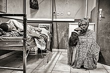
A mother spends days sitting with her son, a hospital patient in Mali

An inpatient (or in-patient), on the other hand, is "admitted" to stay in a hospital overnight or for an indeterminate time, usually, several days or weeks, though in some extreme cases, such as with coma or persistent vegetative state, patients can stay in hospitals for years, sometimes until death. Treatment provided in this fashion is called inpatient care. The admission to the hospital involves the production of an admission note. The leaving of the hospital is officially termed discharge, and involves a corresponding discharge note, and sometimes an assessment process to consider ongoing needs. In the English National Health Service this may take the form of "Discharge to Assess" - where the assessment takes place after the patient has gone home.[2]

Misdiagnosis is the leading cause of medical error in outpatient facilities. When the U.S. Institute of Medicine's groundbreaking 1999 report, To Err Is Human, found up to 98,000 hospital patients die from preventable medical errors in the U.S. each year,[3] early efforts focused on inpatient safety.[4] While patient safety efforts have focused on inpatient hospital settings for more than a decade, medical errors are even more likely to happen in a doctor's office or outpatient clinic or center.[citation needed]

Day patient

[edit]

A day patient (or day-patient) is a patient who is using the full range of services of a hospital or clinic but is not expected to stay the night. The term was originally used by psychiatric hospital services using of this patient type to care for people needing support to make the transition from in-patient to out-patient care. However, the term is now also heavily used for people attending hospitals for day surgery.

Alternative terminology

[edit]

Because of concerns such as dignity, human rights and political correctness, the term "patient" is not always used to refer to a person receiving health care. Other terms that are sometimes used include health consumer, healthcare consumer, customer or client. However, such terminology may be offensive to those receiving public health care, as it implies a business relationship.

In veterinary medicine, the client is the owner or guardian of the patient. These may be used by governmental agencies, insurance companies, patient groups, or health care facilities. Individuals who use or have used psychiatric services may alternatively refer to themselves as consumers, users, or survivors.

In nursing homes and assisted living facilities, the term resident is generally used in lieu of patient.[5] Similarly, those receiving home health care are called clients.

Patient-centered healthcare

[edit]

The doctor–patient relationship has sometimes been characterized as silencing the voice of patients.[6] It is now widely agreed that putting patients at the centre of healthcare[7] by trying to provide a consistent, informative and respectful service to patients will improve both outcomes and patient satisfaction.[8]

When patients are not at the centre of healthcare, when institutional procedures and targets eclipse local concerns, then patient neglect is possible.[9] Incidents, such as the Stafford Hospital scandal, Winterbourne View hospital abuse scandal and the Veterans Health Administration controversy of 2014 have shown the dangers of prioritizing cost control over the patient experience.[10] Investigations into these and other scandals have recommended that healthcare systems put patient experience at the center, and especially that patients themselves are heard loud and clear within health services.[11]

There are many reasons for why health services should listen more to patients. Patients spend more time in healthcare services than regulators or quality controllers, and can recognize problems such as service delays, poor hygiene, and poor conduct.[12] Patients are particularly good at identifying soft problems, such as attitudes, communication, and 'caring neglect',[9] that are difficult to capture with institutional monitoring.[13]

One important way in which patients can be placed at the centre of healthcare is for health services to be more open about patient complaints.[14] Each year many hundreds of thousands of patients complain about the care they have received, and these complaints contain valuable information for any health services which want to learn about and improve patient experience.[15]

See also

[edit]
  • Casualty
  • e-Patient
  • Mature minor doctrine
  • Nurse-client relationship
  • Patient abuse
  • Patient advocacy
  • Patient empowerment
  • Patients' Bill of Rights
  • Radiological protection of patients
  • Therapeutic inertia
  • Virtual patient
  • Patient UK

References

[edit]
  1. ^ Neuberger, J. (1999-06-26). "Do we need a new word for patients?". BMJ: British Medical Journal. 318 (7200): 1756–1758. doi:10.1136/bmj.318.7200.1756. ISSN 0959-8138. PMC 1116090. PMID 10381717.
  2. ^ "Unpaid carers' rights are overlooked in hospital discharge". Health Service Journal. 8 September 2021. Retrieved 16 October 2021.
  3. ^ Institute of Medicine (US) Committee on Quality of Health Care in America; Kohn, L. T.; Corrigan, J. M.; Donaldson, M. S. (2000). Kohn, Linda T.; Corrigan, Janet M.; Donaldson, Molla S. (eds.). To Err Is Human: Building a Safer Health System. Washington D.C.: National Academy Press. doi:10.17226/9728. ISBN 0-309-06837-1. PMID 25077248.
  4. ^ Bates, David W.; Singh, Hardeep (November 2018). "Two Decades Since: An Assessment Of Progress And Emerging Priorities In Patient Safety". Health Affairs. 37 (11): 1736–1743. doi:10.1377/hlthaff.2018.0738. PMID 30395508.
  5. ^ American Red Cross (1993). Foundations for Caregiving. St. Louis: Mosby Lifeline. ISBN 978-0801665158.
  6. ^ Clark, Jack A.; Mishler, Elliot G. (September 1992). "Attending to patients' stories: reframing the clinical task". Sociology of Health and Illness. 14 (3): 344–372. doi:10.1111/1467-9566.ep11357498.
  7. ^ Stewart, M (24 February 2001). "Towards a Global Definition of Patient Centred Care". BMJ. 322 (7284): 444–5. doi:10.1136/bmj.322.7284.444. PMC 1119673. PMID 11222407.
  8. ^ Frampton, Susan B.; Guastello, Sara; Hoy, Libby; Naylor, Mary; Sheridan, Sue; Johnston-Fleece, Michelle (31 January 2017). "Harnessing Evidence and Experience to Change Culture: A Guiding Framework for Patient and Family Engaged Care". NAM Perspectives. 7 (1). doi:10.31478/201701f.
  9. ^ a b Reader, TW; Gillespie, A (30 April 2013). "Patient Neglect in Healthcare Institutions: A Systematic Review and Conceptual Model". BMC Health Serv Res. 13: 156. doi:10.1186/1472-6963-13-156. PMC 3660245. PMID 23631468.
  10. ^ Bloche, MG (17 March 2016). "Scandal as a Sentinel Event--Recognizing Hidden Cost-Quality Trade-offs". N Engl J Med. 374 (11): 1001–3. doi:10.1056/NEJMp1502629. PMID 26981930.
  11. ^ Report of the Mid Staffordshire NHS Foundation Trust Public Inquiry: Executive Summary. London: Stationery Office. 6 February 2013. ISBN 9780102981476. Retrieved 23 June 2020.
  12. ^ Weingart, SN; Pagovich, O; Sands, DZ; Li, JM; Aronson, MD; Davis, RB; Phillips, RS; Bates, DW (April 2006). "Patient-reported Service Quality on a Medicine Unit". Int J Qual Health Care. 18 (2): 95–101. doi:10.1093/intqhc/mzi087. PMID 16282334.
  13. ^ Levtzion-Korach, O; Frankel, A; Alcalai, H; Keohane, C; Orav, J; Graydon-Baker, E; Barnes, J; Gordon, K; Puopulo, AL; Tomov, EI; Sato, L; Bates, DW (September 2010). "Integrating Incident Data From Five Reporting Systems to Assess Patient Safety: Making Sense of the Elephant". Jt Comm J Qual Patient Saf. 36 (9): 402–10. doi:10.1016/s1553-7250(10)36059-4. PMID 20873673.
  14. ^ Berwick, Donald M. (January 2009). "What 'Patient-Centered' Should Mean: Confessions Of An Extremist". Health Affairs. 28 (Supplement 1): w555 – w565. doi:10.1377/hlthaff.28.4.w555. PMID 19454528.
  15. ^ Reader, TW; Gillespie, A; Roberts, J (August 2014). "Patient Complaints in Healthcare Systems: A Systematic Review and Coding Taxonomy". BMJ Qual Saf. 23 (8): 678–89. doi:10.1136/bmjqs-2013-002437. PMC 4112446. PMID 24876289.
[edit]
  • Jadad AR, Rizo CA, Enkin MW (June 2003). "I am a good patient, believe it or not". BMJ. 326 (7402): 1293–5. doi:10.1136/bmj.326.7402.1293. PMC 1126181. PMID 12805157.
    a peer-reviewed article published in the British Medical Journal's (BMJ) first issue dedicated to patients in its 160-year history
  • Sokol DK (21 February 2004). "How (not) to be a good patient". BMJ. 328 (7437): 471. doi:10.1136/bmj.328.7437.471. PMC 344286.
    review article with views on the meaning of the words "good doctor" vs. "good patient"
  • "Time Magazine's Dr. Scott Haig Proves that Patients Need to Be Googlers!" – Mary Shomons response to the Time Magazine article "When the Patient is a Googler"