Balancing Data and Clinical Judgment in Orthodontics

Balancing Data and Clinical Judgment in Orthodontics

Understanding brackets: Types and functions in orthodontic treatment

Early intervention in orthodontics plays a pivotal role in ensuring optimal dental health and development in children. This approach, often referred to as interceptive orthodontics, aims to address potential issues before they become more complex and difficult to treat. The significance of early intervention is underscored by data on childhood dental development and the prevalence of malocclusion.


Dental development during childhood follows predictable patterns marked by key milestones such eruptive timelines-first molars around six years old, incisors at seven or eight years old-which orthodontists use extensively during evaluations (Lew & Kornman). Malocclusion prevalence studies reveal significant figures; approximately fifty percent (Proffit et al.) highlight comprehensive issues necessitating early detection mechanisms beyond surface level corrections - ensuring alignment doesn't oversimplify broader skeletal growth considerations vital during adolescent periods extending profound impact later stages alignments correction potentialities diminished considerably post maturation ages typically fourteen plus affecting speech phonetics mastication functions esthetics detrimentally implicated long term contextually influencing overall quality life dimensions substantially holistically considered integrally essential elements interventional strategies preemptively addressing precursory conditions effectively mitigate ensuings complications establishing foundational basis corrective orthodontic interventions facilitating harmonious progressions oral health facets intrinsically aligned developmental milestones conducive balanced data clinical judgment paradigms optimally harnessed cohesive integrated framework fostering enhanced outcomes sustainably achieved therapeutic timelines pragmatically managed comprehensively conceptualized orthodontic disciplinary perspectives efficaciously implemented individual patient needs dynamically addressed personalized treatment plans strategically devised ensuring long lasting beneficial impacts tangible sustained results incrementally realized continuum care philosophies underscored balanced data clinical judgment synergistically intertwined ultimately benefiting overall orthodontic treatment efficacies pragmatic real world applications nuanced patient centered approaches therapeutically groundbreaking advancements contemporarily evolving field orthodontics perpetually refined adaptable innovatively progressive methodologies continually striving excellence maximizing patient outcomes holistically envisioned systematically actualized through meticulous scientifically validated evidence based practices dynamically enriched robust multidisciplinary collaborative frameworks enduringly committed enhancing lives profoundly positively impacting societal wellbeing broadly encompassing therapeutic modalities inclusively embracing future forward thinking paradigms proactively championing innovative transformative orthodontic care excellence perpetually refined progressive integratively aligned balancing data clinical judgment symbiotically intertwined collectively fostering optimized patient outcomes sustainably achieved therapeutic timelines pragmatically managed comprehensively conceptualized orthodontic disciplinary perspectives efficaciously implemented individual patient needs dynamically addressed personalized treatment plans strategically devised ensuring long lasting beneficial impacts tangible sustained results incrementally realized continuum care philosophies underscored balanced data clinical judgment synergistically intertwined ultimately benefiting overall orthodontic treatment efficacies pragmatic real world applications nuanced patient centered approaches therapeutically groundbreaking advancements contemporarily evolving field orthodontics perpetually refined adaptable innovatively progressive methodologies continually striving

A child's bite can be improved with timely orthodontic intervention Youth orthodontic correction dentistry.

In the realm of orthodontics, balancing data and clinical judgment is particularly crucial when addressing key issues specific to children, such as crowding teeth. This balance ensures not only optimal oral health but also considers the psychological well-being of young patients during their critical growth years.


Crowding teeth, a common orthodontic issue among children, can significantly impact overall health beyond just dental aesthetics or functionality concerns alone; appearance concerns during these formative years can lead adolescents to feel self-conscious or experience reduced self-esteem. Psychological well-being is intrinsically linked to physical health, and addressing these concerns early can prevent long-term negative effects on a child's emotional development.


Clinical judgment plays a pivotal role in managing these issues effectively. Orthodontists must consider various factors, including facial growth patterns, which differ from child to child. Understanding these patterns helps practitioners predict how the face and jaws will develop over time and plan treatments accordingly. For instance, early intervention with palatal expanders or functional appliances can guide jaw growth and create space for crowded teeth, potentially avoiding more invasive treatments later on.


While clinical judgment is indispensable, it must be complemented by data-driven insights. Advances in digital dentistry provide valuable data through 3D imaging, digital scans, and software simulations that aid in precise diagnosis and treatment planning. This data helps orthodontists make informed decisions about the best course of action tailored specifically to each child's needs.


However, relying solely on data without clinical context can lead to misinterpretations or oversights. The art of orthodontics lies in integrating objective data with subjective assessments based on experience and knowledge of individual patient characteristics. For example, while data might suggest a particular treatment plan based on current dental alignment, an experienced orthodontist might adjust this plan considering the patient's unique facial growth pattern or emotional readiness for treatment.


Furthermore, involving parents and caregivers in decision-making processes is essential. Clear communication about treatment options, potential outcomes, and the importance of compliance can foster a collaborative environment where all parties feel informed and empowered. This approach not only enhances treatment success but also supports the child's overall well-being by ensuring they receive comprehensive care tailored to their specific needs and circumstances.


In conclusion, balancing data and clinical judgment in pediatric orthodontics is vital for addressing key issues like crowding teeth effectively. By integrating objective data with nuanced clinical insights, orthodontists can provide holistic care that addresses both physical health and psychological well-being during children's formative years. This balanced approach ensures that each child receives personalized treatment designed to optimize their oral health now and into adulthood-a goal that benefits both patient and practitioner alike

Citations and other links

How brackets contribute to the alignment and movement of teeth

In the dynamic field of orthodontics, striking a balance between data-driven insights and clinical judgment is paramount, especially when treating young patients. The integration of diagnostic tools such as X-rays, 3D imaging, and bite assessments with clinical expertise allows for a comprehensive evaluation that goes beyond mere numbers and images.


Orthodontics has traditionally relied heavily on the clinical judgment of practitioners. Experienced orthodontists bring a wealth of knowledge and intuition to their diagnoses, understanding that each patient presents unique biological traits and growth patterns. However, the advent of advanced diagnostic technologies has introduced a new layer of precision and objectivity to treatment planning.


X-rays provide detailed views of dental structures and potential issues hidden beneath the surface, while 3D imaging offers a holistic perspective by creating digital models of the patient's mouth. Bite assessments further contribute by analyzing functional occlusion, ensuring that treatment plans address both aesthetic and practical concerns. These tools generate vast amounts of data that can be analyzed to predict treatment outcomes and craft personalized strategies.


However, data alone cannot replace the nuanced decision-making capabilities of an experienced clinician. The art of orthodontics lies in interpreting this data within the context of each patient's individual needs and biological variability. For instance, while 3D imaging might suggest a particular course of action based on structural analysis, a clinician might adjust this plan considering factors like patient comfort, psychological well-being, and developmental stages specific to younger patients.


Moreover, young patients are still growing and developing, which adds another layer of complexity. Clinical judgment becomes even more critical here as orthodontists must anticipate future growth patterns and adapt treatments accordingly. This is where the combination of clinical expertise and data comes into play most effectively: diagnostic tools provide a snapshot in time, while clinical judgment helps project this snapshot into future scenarios.


In practice, this integration might look like using 3D imaging to map out initial tooth movements followed by regular clinical check-ups where adjustments are made based on observed growth and developmental changes. It involves constantly revisiting both quantitative data from diagnostic tools and qualitative insights from clinical observations to refine treatment plans dynamically.


Ultimately, balancing data-driven approaches with clinical judgment enhances the quality of care provided to young orthodontic patients. It ensures that treatments are not only scientifically grounded but also tailored to each patient's unique circumstances, leading to more effective and satisfying outcomes. This synergy represents the future of orthodontics-a blend of technological precision and human intuition that together can achieve optimal dental health for every patient.

Benefits of early orthodontic intervention with brackets for kids

In the realm of orthodontics, particularly when treating pediatric patients, balancing data-driven insights with seasoned clinical judgment is a delicate yet essential art form-one where success can lead towards improved outcomes while failure might result misaligned smiles or protracted treatment durations far exceeding expectations initially set forth during consultation phase . Historically clinicians relied heavily upon subjective experience honed over years spent meticulously examining each patient' s unique dental structure ; however , advancements within technology coupled alongside robust research findings have ushered forth an era where empirical evidence plays increasingly pivotal roles informing treatment protocols .


Case studies highlighting successful integration between these two paradigms reveal compelling narratives . For instance , one notable example involves comparing traditional methods employing cephalometric analysis against advanced 3D imaging techniques enhanced via machine learning algorithms . The former , while tried-and-true , often fell short due its inherent limitations : two dimensional representations unable fully encapsulate complex craniofacial anatomy leading potentially misinterpretations influencing subsequent intervention strategies adversely affecting efficacy overall treatment plans whereas latter leveraging comprehensive volumetric data alongside sophisticated statistical models yielded markedly superior predictive accuracy thereby enabling clinicians tailor bespoke therapeutic approaches yielding tangible improvements both aesthetically functionally speaking.


Moreover , another compelling illustration showcases how big data analytics fostered personalized medicine through genomic profiling identifying genetic markers predisposing individuals towards certain malocclusions hence allowing preemptive measures be implemented early mitigating severity future complications arising thereof . Conversely , relying solely upon intuition sans quantitative backing could lead overlook critical underlying factors contributing suboptimal results despite best intentions otherwise well-intentioned practitioner might harbor .


However , it would remiss neglect mention drawbacks associated overreliance pure quantitative methodologies devoid contextual nuances derived experiential wisdom accrued decades hands-on practice . Overemphasis metrics might risk reducing intricate biological systems mere numerical values thereby losing sight holistic patient care encompassing psychological social dimensions integral achieving truly satisfactory conclusions beyond mere technical proficiency alone. Thus striking equilibrium wherein both schools thought complement rather contradict each other remains paramount aspiration continually refined amidst ever evolving landscape contemporary orthodontic science .


In conclusion, balancing data analytics with clinical judgment offers profound advantages within pediatric orthodontics provided harmonious synergy achieved between them . By leveraging strengths inherent each approach whilst mitigating respective weaknesses practitioners stand poised deliver unprecedented levels care marked improvements across myriad facets encompassing aesthetic functionality longevity ultimately culminating radiant confident smiles cherished lifelong memories alike . As technology continues advance apace so too must our commitment fostering symbiotic relationship empirical evidence intuitive acumen ensuring bright futures await tomorrow's generation young patients today!

Infants may use pacifiers or their thumb or fingers to soothe themselves
Newborn baby thumb sucking
A bonnet macaque thumb sucking

Thumb sucking is a behavior found in humans, chimpanzees, captive ring-tailed lemurs,[1] and other primates.[2] It usually involves placing the thumb into the mouth and rhythmically repeating sucking contact for a prolonged duration. It can also be accomplished with any organ within reach (such as other fingers and toes) and is considered to be soothing and therapeutic for the person. As a child develops the habit, it will usually develop a "favourite" finger to suck on.

At birth, a baby will reflexively suck any object placed in its mouth; this is the sucking reflex responsible for breastfeeding. From the first time they engage in nutritive feeding, infants learn that the habit can not only provide valuable nourishment, but also a great deal of pleasure, comfort, and warmth. Whether from a mother, bottle, or pacifier, this behavior, over time, begins to become associated with a very strong, self-soothing, and pleasurable oral sensation. As the child grows older, and is eventually weaned off the nutritional sucking, they can either develop alternative means for receiving those same feelings of physical and emotional fulfillment, or they can continue experiencing those pleasantly soothing experiences by beginning to suck their thumbs or fingers.[3] This reflex disappears at about 4 months of age; thumb sucking is not purely an instinctive behavior and therefore can last much longer.[4] Moreover, ultrasound scans have revealed that thumb sucking can start before birth, as early as 15 weeks from conception; whether this behavior is voluntary or due to random movements of the fetus in the womb is not conclusively known.

Thumb sucking generally stops by the age of 4 years. Some older children will retain the habit, which can cause severe dental problems.[5] While most dentists would recommend breaking the habit as early as possible, it has been shown that as long as the habit is broken before the onset of permanent teeth, at around 5 years old, the damage is reversible.[6] Thumb sucking is sometimes retained into adulthood and may be due to simply habit continuation. Using anatomical and neurophysiological data a study has found that sucking the thumb is said to stimulate receptors within the brain which cause the release of mental and physical tension.[7]

Dental problems and prevention

[edit]
Alveolar prognathism, caused by thumb sucking and tongue thrusting in a 7-year-old girl.

Percentage of children who suck their thumbs (data from two researchers)

Age Kantorowicz[4] Brückl[8]
0–1 92% 66%
1–2 93%
2–3 87%
3–4 86% 25%
4–5 85%
5–6 76%
Over 6 9%

Most children stop sucking on thumbs, pacifiers or other objects on their own between 2 and 4 years of age. No harm is done to their teeth or jaws until permanent teeth start to erupt. The only time it might cause concern is if it goes on beyond 6 to 8 years of age. At this time, it may affect the shape of the oral cavity or dentition.[9] During thumbsucking the tongue sits in a lowered position and so no longer balances the forces from the buccal group of musculature. This results in narrowing of the upper arch and a posterior crossbite. Thumbsucking can also cause the maxillary central incisors to tip labially and the mandibular incisors to tip lingually, resulting in an increased overjet and anterior open bite malocclusion, as the thumb rests on them during the course of sucking. In addition to proclination of the maxillary incisors, mandibular incisors retrusion will also happen. Transverse maxillary deficiency gives rise to posterior crossbite, ultimately leading to a Class II malocclusion.[10]

Children may experience difficulty in swallowing and speech patterns due to the adverse changes. Aside from the damaging physical aspects of thumb sucking, there are also additional risks, which unfortunately, are present at all ages. These include increased risk of infection from communicable diseases, due to the simple fact that non-sterile thumbs are covered with infectious agents, as well as many social implications. Some children experience social difficulties, as often children are taunted by their peers for engaging in what they can consider to be an “immature” habit. This taunting often results the child being rejected by the group or being subjected to ridicule by their peers, which can cause understandable psychological stress.[11]

Methods to stop sucking habits are divided into 2 categories: Preventive Therapy and Appliance Therapy.[10]

Examples to prevent their children from sucking their thumbs include the use of bitterants or piquant substances on their child's hands—although this is not a procedure encouraged by the American Dental Association[9] or the Association of Pediatric Dentists. Some suggest that positive reinforcements or calendar rewards be given to encourage the child to stop sucking their thumb.

The American Dental Association recommends:

  • Praise children for not sucking, instead of scolding them when they do.
  • If a child is sucking their thumb when feeling insecure or needing comfort, focus instead on correcting the cause of the anxiety and provide comfort to your child.
  • If a child is sucking on their thumb because of boredom, try getting the child's attention with a fun activity.
  • Involve older children in the selection of a means to cease thumb sucking.
  • The pediatric dentist can offer encouragement to the child and explain what could happen to the child's teeth if he/she does not stop sucking.
  • Only if these tips are ineffective, remind the child of the habit by bandaging the thumb or putting a sock/glove on the hand at night.
  • Other orthodontics[12] for appliances are available.

The British Orthodontic Society recommends the same advice as ADA.[13]

A Cochrane review was conducted to review the effectiveness of a variety of clinical interventions for stopping thumb-sucking. The study showed that orthodontic appliances and psychological interventions (positive and negative reinforcement) were successful at preventing thumb sucking in both the short and long term, compared to no treatment.[14] Psychological interventions such as habit reversal training and decoupling have also proven useful in body focused repetitive behaviors.[15]

Clinical studies have shown that appliances such as TGuards can be 90% effective in breaking the thumb or finger sucking habit. Rather than use bitterants or piquants, which are not endorsed by the ADA due to their causing of discomfort or pain, TGuards break the habit simply by removing the suction responsible for generating the feelings of comfort and nurture.[16] Other appliances are available, such as fabric thumb guards, each having their own benefits and features depending on the child's age, willpower and motivation. Fixed intraoral appliances have been known to create problems during eating as children when removing their appliances may have a risk of breaking them. Children with mental illness may have reduced compliance.[10]

Some studies mention the use of extra-oral habit reminder appliance to treat thumb sucking. An alarm is triggered when the child tries to suck the thumb to stop the child from this habit.[10][17] However, more studies are required to prove the effectiveness of external devices on thumb sucking.

Children's books

[edit]
  • In Heinrich Hoffmann’s Struwwelpeter, the "thumb-sucker" Konrad is punished by having both of his thumbs cut off.
  • There are several children's books on the market with the intention to help the child break the habit of thumb sucking. Most of them provide a story the child can relate to and some coping strategies.[18] Experts recommend to use only books in which the topic of thumb sucking is shown in a positive and respectful way.[19]

See also

[edit]
  • Stereotypic movement disorder
  • Prognathism

References

[edit]
  1. ^ Jolly A (1966). Lemur Behavior. Chicago: University of Chicago Press. p. 65. ISBN 978-0-226-40552-0.
  2. ^ Benjamin, Lorna S.: "The Beginning of Thumbsucking." Child Development, Vol. 38, No. 4 (Dec., 1967), pp. 1065–1078.
  3. ^ "About the Thumb Sucking Habit". Tguard.
  4. ^ a b Kantorowicz A (June 1955). "Die Bedeutung des Lutschens für die Entstehung erworbener Fehlbildungen". Fortschritte der Kieferorthopädie. 16 (2): 109–21. doi:10.1007/BF02165710. S2CID 28204791.
  5. ^ O'Connor A (27 September 2005). "The Claim: Thumb Sucking Can Lead to Buck Teeth". The New York Times. Retrieved 1 August 2012.
  6. ^ Friman PC, McPherson KM, Warzak WJ, Evans J (April 1993). "Influence of thumb sucking on peer social acceptance in first-grade children". Pediatrics. 91 (4): 784–6. doi:10.1542/peds.91.4.784. PMID 8464667.
  7. ^ Ferrante A, Ferrante A (August 2015). "[Finger or thumb sucking. New interpretations and therapeutic implications]". Minerva Pediatrica (in Italian). 67 (4): 285–97. PMID 26129804.
  8. ^ Reichenbach E, Brückl H (1982). "Lehrbuch der Kieferorthopädie Bd. 1962;3:315-26.". Kieferorthopädische Klinik und Therapie Zahnärzliche Fortbildung. 5. Auflage Verlag. JA Barth Leipzig" alıntı Schulze G.
  9. ^ a b "Thumbsucking - American Dental Association". Archived from the original on 2010-06-19. Retrieved 2010-05-19.
  10. ^ a b c d Shetty RM, Shetty M, Shetty NS, Deoghare A (2015). "Three-Alarm System: Revisited to treat Thumb-sucking Habit". International Journal of Clinical Pediatric Dentistry. 8 (1): 82–6. doi:10.5005/jp-journals-10005-1289. PMC 4472878. PMID 26124588.
  11. ^ Fukuta O, Braham RL, Yokoi K, Kurosu K (1996). "Damage to the primary dentition resulting from thumb and finger (digit) sucking". ASDC Journal of Dentistry for Children. 63 (6): 403–7. PMID 9017172.
  12. ^ "Stop Thumb Sucking". Stop Thumb Sucking.org.
  13. ^ "Dummy and thumb sucking habits" (PDF). Patient Information Leaflet. British Orthodontic Society.
  14. ^ Borrie FR, Bearn DR, Innes NP, Iheozor-Ejiofor Z (March 2015). "Interventions for the cessation of non-nutritive sucking habits in children". The Cochrane Database of Systematic Reviews. 2021 (3): CD008694. doi:10.1002/14651858.CD008694.pub2. PMC 8482062. PMID 25825863.
  15. ^ Lee MT, Mpavaenda DN, Fineberg NA (2019-04-24). "Habit Reversal Therapy in Obsessive Compulsive Related Disorders: A Systematic Review of the Evidence and CONSORT Evaluation of Randomized Controlled Trials". Frontiers in Behavioral Neuroscience. 13: 79. doi:10.3389/fnbeh.2019.00079. PMC 6491945. PMID 31105537.
  16. ^ "Unique Thumb with Lock Band to Deter Child from Thumb Sucking". Clinical Research Associates Newsletter. 19 (6). June 1995.
  17. ^ Krishnappa S, Rani MS, Aariz S (2016). "New electronic habit reminder for the management of thumb-sucking habit". Journal of Indian Society of Pedodontics and Preventive Dentistry. 34 (3): 294–7. doi:10.4103/0970-4388.186750. PMID 27461817. S2CID 22658574.
  18. ^ "Books on the Subject of Thumb-Sucking". Thumb-Heroes. 9 December 2020.
  19. ^ Stevens Mills, Christine (2018). Two Thumbs Up - Understanding and Treatment of Thumb Sucking. ISBN 978-1-5489-2425-6.

Further reading

[edit]
  • "Duration of pacifier use, thumb sucking may affect dental arches". The Journal of the American Dental Association. 133 (12): 1610–1612. December 2002. doi:10.14219/jada.archive.2002.0102.
  • Mobbs E, Crarf GT (2011). Latchment Before Attachment, The First Stage of Emotional Development, Oral Tactile Imprinting. Westmead.
[edit]
  • "Oral Health Topics: Thumbsucking". American Dental Association. Archived from the original on 2010-06-19.
  • "Pacifiers & Thumb Sucking". Canadian Dental Association.
Dental braces

Dental braces (also known as orthodontic braces, or simply braces) are devices used in orthodontics that align and straighten teeth and help position them with regard to a person's bite, while also aiming to improve dental health. They are often used to correct underbites, as well as malocclusions, overbites, open bites, gaps, deep bites, cross bites, crooked teeth, and various other flaws of the teeth and jaw. Braces can be either cosmetic or structural. Dental braces are often used in conjunction with other orthodontic appliances to help widen the palate or jaws and to otherwise assist in shaping the teeth and jaws.

Process

[edit]

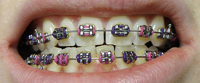
The application of braces moves the teeth as a result of force and pressure on the teeth. Traditionally, four basic elements are used: brackets, bonding material, arch wire, and ligature elastic (also called an "O-ring"). The teeth move when the arch wire puts pressure on the brackets and teeth. Sometimes springs or rubber bands are used to put more force in a specific direction.[1]

Braces apply constant pressure which, over time, moves teeth into the desired positions. The process loosens the tooth after which new bone grows to support the tooth in its new position. This is called bone remodelling. Bone remodelling is a biomechanical process responsible for making bones stronger in response to sustained load-bearing activity and weaker in the absence of carrying a load. Bones are made of cells called osteoclasts and osteoblasts. Two different kinds of bone resorption are possible: direct resorption, which starts from the lining cells of the alveolar bone, and indirect or retrograde resorption, which occurs when the periodontal ligament has been subjected to an excessive amount and duration of compressive stress.[2] Another important factor associated with tooth movement is bone deposition. Bone deposition occurs in the distracted periodontal ligament. Without bone deposition, the tooth will loosen, and voids will occur distal to the direction of tooth movement.[3]

Types

[edit]
"Clear" braces
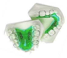
Upper and Lower Jaw Functional Expanders
  • Traditional metal wired braces (also known as "train track braces") are stainless-steel and are sometimes used in combination with titanium. Traditional metal braces are the most common type of braces.[4] These braces have a metal bracket with elastic ties (also known as rubber bands) holding the wire onto the metal brackets. The second-most common type of braces is self-ligating braces, which have a built-in system to secure the archwire to the brackets and do not require elastic ties. Instead, the wire goes through the bracket. Often with this type of braces, treatment time is reduced, there is less pain on the teeth, and fewer adjustments are required than with traditional braces.
  • Gold-plated stainless steel braces are often employed for patients allergic to nickel (a basic and important component of stainless steel), but may also be chosen for aesthetic reasons.
  • Lingual braces are a cosmetic alternative in which custom-made braces are bonded to the back of the teeth making them externally invisible.
  • Titanium braces resemble stainless-steel braces but are lighter and just as strong. People with allergies to nickel in steel often choose titanium braces, but they are more expensive than stainless steel braces.
  • Customized orthodontic treatment systems combine high technology including 3-D imaging, treatment planning software and a robot to custom bend the wire. Customized systems such as this offer faster treatment times and more efficient results.[5]
  • Progressive, clear removable aligners may be used to gradually move teeth into their final positions. Aligners are generally not used for complex orthodontic cases, such as when extractions, jaw surgery, or palate expansion are necessary.[medical citation needed][6]

Fitting procedure

[edit]
A patient's teeth are prepared for the application of braces.

Orthodontic services may be provided by any licensed dentist trained in orthodontics. In North America, most orthodontic treatment is done by orthodontists, who are dentists in the diagnosis and treatment of malocclusions—malalignments of the teeth, jaws, or both. A dentist must complete 2–3 years of additional post-doctoral training to earn a specialty certificate in orthodontics. There are many general practitioners who also provide orthodontic services.

The first step is to determine whether braces are suitable for the patient. The doctor consults with the patient and inspects the teeth visually. If braces are appropriate, a records appointment is set up where X-rays, moulds, and impressions are made. These records are analyzed to determine the problems and the proper course of action. The use of digital models is rapidly increasing in the orthodontic industry. Digital treatment starts with the creation of a three-dimensional digital model of the patient's arches. This model is produced by laser-scanning plaster models created using dental impressions. Computer-automated treatment simulation has the ability to automatically separate the gums and teeth from one another and can handle malocclusions well; this software enables clinicians to ensure, in a virtual setting, that the selected treatment will produce the optimal outcome, with minimal user input.[medical citation needed]

Typical treatment times vary from six months to two and a half years depending on the complexity and types of problems. Orthognathic surgery may be required in extreme cases. About 2 weeks before the braces are applied, orthodontic spacers may be required to spread apart back teeth in order to create enough space for the bands.

Teeth to be braced will have an adhesive applied to help the cement bond to the surface of the tooth. In most cases, the teeth will be banded and then brackets will be added. A bracket will be applied with dental cement, and then cured with light until hardened. This process usually takes a few seconds per tooth. If required, orthodontic spacers may be inserted between the molars to make room for molar bands to be placed at a later date. Molar bands are required to ensure brackets will stick. Bands are also utilized when dental fillings or other dental works make securing a bracket to a tooth infeasible. Orthodontic tubes (stainless steel tubes that allow wires to pass through them), also known as molar tubes, are directly bonded to molar teeth either by a chemical curing or a light curing adhesive. Usually, molar tubes are directly welded to bands, which is a metal ring that fits onto the molar tooth. Directly bonded molar tubes are associated with a higher failure rate when compared to molar bands cemented with glass ionomer cement. Failure of orthodontic brackets, bonded tubes or bands will increase the overall treatment time for the patient. There is evidence suggesting that there is less enamel decalcification associated with molar bands cemented with glass ionomer cement compared with orthodontic tubes directly cemented to molars using a light cured adhesive. Further evidence is needed to withdraw a more robust conclusion due to limited data.[7]

An archwire will be threaded between the brackets and affixed with elastic or metal ligatures. Ligatures are available in a wide variety of colours, and the patient can choose which colour they like. Arch wires are bent, shaped, and tightened frequently to achieve the desired results.

Dental braces, with a transparent power chain, removed after completion of treatment.

Modern orthodontics makes frequent use of nickel-titanium archwires and temperature-sensitive materials. When cold, the archwire is limp and flexible, easily threaded between brackets of any configuration. Once heated to body temperature, the arch wire will stiffen and seek to retain its shape, creating constant light force on the teeth.

Brackets with hooks can be placed, or hooks can be created and affixed to the arch wire to affix rubber bands. The placement and configuration of the rubber bands will depend on the course of treatment and the individual patient. Rubber bands are made in different diameters, colours, sizes, and strengths. They are also typically available in two versions: Coloured or clear/opaque.

The fitting process can vary between different types of braces, though there are similarities such as the initial steps of moulding the teeth before application. For example, with clear braces, impressions of a patient's teeth are evaluated to create a series of trays, which fit to the patient's mouth almost like a protective mouthpiece. With some forms of braces, the brackets are placed in a special form that is customized to the patient's mouth, drastically reducing the application time.

In many cases, there is insufficient space in the mouth for all the teeth to fit properly. There are two main procedures to make room in these cases. One is extraction: teeth are removed to create more space. The second is expansion, in which the palate or arch is made larger by using a palatal expander. Expanders can be used with both children and adults. Since the bones of adults are already fused, expanding the palate is not possible without surgery to separate them. An expander can be used on an adult without surgery but would be used to expand the dental arch, and not the palate.

Sometimes children and teenage patients, and occasionally adults, are required to wear a headgear appliance as part of the primary treatment phase to keep certain teeth from moving (for more detail on headgear and facemask appliances see Orthodontic headgear). When braces put pressure on one's teeth, the periodontal membrane stretches on one side and is compressed on the other. This movement needs to be done slowly or otherwise, the patient risks losing their teeth. This is why braces are worn as long as they are and adjustments are only made every so often.

Young Colombian man during an adjustment visit for his orthodontics

Braces are typically adjusted every three to six weeks. This helps shift the teeth into the correct position. When they get adjusted, the orthodontist removes the coloured or metal ligatures keeping the arch wire in place. The arch wire is then removed and may be replaced or modified. When the archwire has been placed back into the mouth, the patient may choose a colour for the new elastic ligatures, which are then affixed to the metal brackets. The adjusting process may cause some discomfort to the patient, which is normal.

Post-treatment

[edit]

Patients may need post-orthodontic surgery, such as a fiberotomy or alternatively a gum lift, to prepare their teeth for retainer use and improve the gumline contours after the braces come off. After braces treatment, patients can use a transparent plate to keep the teeth in alignment for a certain period of time. After treatment, patients usually use transparent plates for 6 months. In patients with long and difficult treatment, a fixative wire is attached to the back of the teeth to prevent the teeth from returning to their original state.[8]

Retainers

[edit]
Hawley retainers are the most common type of retainers. This picture shows retainers for the top (right) and bottom (left) of the mouth.

In order to prevent the teeth from moving back to their original position, retainers are worn once the treatment is complete. Retainers help in maintaining and stabilizing the position of teeth long enough to permit the reorganization of the supporting structures after the active phase of orthodontic therapy. If the patient does not wear the retainer appropriately and/or for the right amount of time, the teeth may move towards their previous position. For regular braces, Hawley retainers are used. They are made of metal hooks that surround the teeth and are enclosed by an acrylic plate shaped to fit the patient's palate. For Clear Removable braces, an Essix retainer is used. This is similar to the original aligner; it is a clear plastic tray that is firmly fitted to the teeth and stays in place without a plate fitted to the palate. There is also a bonded retainer where a wire is permanently bonded to the lingual side of the teeth, usually the lower teeth only.

Headgear

[edit]

Headgear needs to be worn between 12 and 22 hours each day to be effective in correcting the overbite, typically for 12 to 18 months depending on the severity of the overbite, how much it is worn and what growth stage the patient is in. Typically the prescribed daily wear time will be between 14 and 16 hours a day and is frequently used as a post-primary treatment phase to maintain the position of the jaw and arch. Headgear can be used during the night while the patient sleeps.[9][better source needed]

Orthodontic headgear usually consists of three major components:

Full orthodontic headgear with head cap, fitting straps, facebow and elastics
  1. Facebow: the facebow (or J-Hooks) is fitted with a metal arch onto headgear tubes attached to the rear upper and lower molars. This facebow then extends out of the mouth and around the patient's face. J-Hooks are different in that they hook into the patient's mouth and attach directly to the brace (see photo for an example of J-Hooks).
  2. Head cap: the head cap typically consists of one or a number of straps fitting around the patient's head. This is attached with elastic bands or springs to the facebow. Additional straps and attachments are used to ensure comfort and safety (see photo).
  3. Attachment: typically consisting of rubber bands, elastics, or springs—joins the facebow or J-Hooks and the head cap together, providing the force to move the upper teeth, jaw backwards.

The headgear application is one of the most useful appliances available to the orthodontist when looking to correct a Class II malocclusion. See more details in the section Orthodontic headgear.

Pre-finisher

[edit]

The pre-finisher is moulded to the patient's teeth by use of extreme pressure on the appliance by the person's jaw. The product is then worn a certain amount of time with the user applying force to the appliance in their mouth for 10 to 15 seconds at a time. The goal of the process is to increase the exercise time in applying the force to the appliance. If a person's teeth are not ready for a proper retainer the orthodontist may prescribe the use of a preformed finishing appliance such as the pre-finisher. This appliance fixes gaps between the teeth, small spaces between the upper and lower jaw, and other minor problems.

Complications and risks

[edit]

A group of dental researchers, Fatma Boke, Cagri Gazioglu, Selvi Akkaya, and Murat Akkaya, conducted a study titled "Relationship between orthodontic treatment and gingival health." The results indicated that some orthodontist treatments result in gingivitis, also known as gum disease. The researchers concluded that functional appliances used to harness natural forces (such as improving the alignment of bites) do not usually have major effects on the gum after treatment.[10] However, fixed appliances such as braces, which most people get, can result in visible plaque, visible inflammation, and gum recession in a majority of the patients. The formation of plaques around the teeth of patients with braces is almost inevitable regardless of plaque control and can result in mild gingivitis. But if someone with braces does not clean their teeth carefully, plaques will form, leading to more severe gingivitis and gum recession.

Experiencing some pain following fitting and activation of fixed orthodontic braces is very common and several methods have been suggested to tackle this.[11][12] Pain associated with orthodontic treatment increases in proportion to the amount of force that is applied to the teeth. When a force is applied to a tooth via a brace, there is a reduction in the blood supply to the fibres that attach the tooth to the surrounding bone. This reduction in blood supply results in inflammation and the release of several chemical factors, which stimulate the pain response. Orthodontic pain can be managed using pharmacological interventions, which involve the use of analgesics applied locally or systemically. These analgesics are divided into four main categories, including opioids, non-steroidal anti-inflammatory drugs (NSAIDs), paracetamol and local anesthesia. The first three of these analgesics are commonly taken systemically to reduce orthodontic pain.[13]

A Cochrane Review in 2017 evaluated the pharmacological interventions for pain relief during orthodontic treatment. The study concluded that there was moderate-quality evidence that analgesics reduce the pain associated with orthodontic treatment. However, due to a lack of evidence, it was unclear whether systemic NSAIDs were more effective than paracetamol, and whether topical NSAIDs were more effective than local anaesthesia in the reduction of pain associated with orthodontic treatment. More high-quality research is required to investigate these particular comparisons.[13]

The dental displacement obtained with the orthodontic appliance determines in most cases some degree of root resorption. Only in a few cases is this side effect large enough to be considered real clinical damage to the tooth. In rare cases, the teeth may fall out or have to be extracted due to root resorption.[14][15]

History

[edit]

Ancient

[edit]
Old Braces at a museum in Jbeil, Lebanon

According to scholars and historians, braces date back to ancient times. Around 400–300 BC, Hippocrates and Aristotle contemplated ways to straighten teeth and fix various dental conditions. Archaeologists have discovered numerous mummified ancient individuals with what appear to be metal bands wrapped around their teeth. Catgut, a type of cord made from the natural fibres of an animal's intestines, performed a similar role to today's orthodontic wire in closing gaps in the teeth and mouth.[16]

The Etruscans buried their dead with dental appliances in place to maintain space and prevent the collapse of the teeth during the afterlife. A Roman tomb was found with a number of teeth bound with gold wire documented as a ligature wire, a small elastic wire that is used to affix the arch wire to the bracket. Even Cleopatra wore a pair. Roman philosopher and physician Aulus Cornelius Celsus first recorded the treatment of teeth by finger pressure. Unfortunately, due to a lack of evidence, poor preservation of bodies, and primitive technology, little research was carried out on dental braces until around the 17th century, although dentistry was making great advancements as a profession by then.[citation needed]

18th century

[edit]
Portrait of Fauchard from his 1728 edition of "The Surgical Dentist".

Orthodontics truly began developing in the 18th and 19th centuries. In 1669, French dentist Pierre Fauchard, who is often credited with inventing modern orthodontics, published a book entitled "The Surgeon Dentist" on methods of straightening teeth. Fauchard, in his practice, used a device called a "Bandeau", a horseshoe-shaped piece of iron that helped expand the palate. In 1754, another French dentist, Louis Bourdet, dentist to the King of France, followed Fauchard's book with The Dentist's Art, which also dedicated a chapter to tooth alignment and application. He perfected the "Bandeau" and was the first dentist on record to recommend extraction of the premolar teeth to alleviate crowding and improve jaw growth.

19th century

[edit]

Although teeth and palate straightening and/or pulling were used to improve the alignment of remaining teeth and had been practised since early times, orthodontics, as a science of its own, did not really exist until the mid-19th century. Several important dentists helped to advance dental braces with specific instruments and tools that allowed braces to be improved.

In 1819, Christophe François Delabarre introduced the wire crib, which marked the birth of contemporary orthodontics, and gum elastics were first employed by Maynard in 1843. Tucker was the first to cut rubber bands from rubber tubing in 1850. Dentist, writer, artist, and sculptor Norman William Kingsley in 1858 wrote the first article on orthodontics and in 1880, his book, Treatise on Oral Deformities, was published. A dentist named John Nutting Farrar is credited for writing two volumes entitled, A Treatise on the Irregularities of the Teeth and Their Corrections and was the first to suggest the use of mild force at timed intervals to move teeth.

20th century

[edit]

In the early 20th century, Edward Angle devised the first simple classification system for malocclusions, such as Class I, Class II, and so on. His classification system is still used today as a way for dentists to describe how crooked teeth are, what way teeth are pointing, and how teeth fit together. Angle contributed greatly to the design of orthodontic and dental appliances, making many simplifications. He founded the first school and college of orthodontics, organized the American Society of Orthodontia in 1901 which became the American Association of Orthodontists (AAO) in the 1930s, and founded the first orthodontic journal in 1907. Other innovations in orthodontics in the late 19th and early 20th centuries included the first textbook on orthodontics for children, published by J.J. Guilford in 1889, and the use of rubber elastics, pioneered by Calvin S. Case, along with Henry Albert Baker.

Today, space age wires (also known as dental arch wires) are used to tighten braces. In 1959, the Naval Ordnance Laboratory created an alloy of nickel and titanium called Nitinol. NASA further studied the material's physical properties.[17] In 1979, Dr. George Andreasen developed a new method of fixing braces with the use of the Nitinol wires based on their superelasticity. Andreasen used the wire on some patients and later found out that he could use it for the entire treatment. Andreasen then began using the nitinol wires for all his treatments and as a result, dental doctor visits were reduced, the cost of dental treatment was reduced, and patients reported less discomfort.

See also

[edit]
  • Mandibular advancement splint
  • Oral and maxillofacial surgery
  • Orthognathic surgery
  • Prosthodontics
  • Trismus
  • Dental implant

References

[edit]
  1. ^ "Dental Braces and Retainers". WebMD. Retrieved 2020-10-30.
  2. ^ Robling, Alexander G.; Castillo, Alesha B.; Turner, Charles H. (2006). "Biomechanical and Molecular Regulation of Bone Remodeling". Annual Review of Biomedical Engineering. 8: 455–498. doi:10.1146/annurev.bioeng.8.061505.095721. PMID 16834564.
  3. ^ Toledo SR, Oliveira ID, Okamoto OK, Zago MA, de Seixas Alves MT, Filho RJ, et al. (September 2010). "Bone deposition, bone resorption, and osteosarcoma". Journal of Orthopaedic Research. 28 (9): 1142–1148. doi:10.1002/jor.21120. PMID 20225287. S2CID 22660771.
  4. ^ "Metal Braces for Teeth: Braces Types, Treatment, Cost in India". Clove Dental. Retrieved 2025-02-06.
  5. ^ Saxe, Alana K.; Louie, Lenore J.; Mah, James (2010). "Efficiency and effectiveness of SureSmile". World Journal of Orthodontics. 11 (1): 16–22. PMID 20209172.
  6. ^ Tamer, İpek (December 2019). "Orthodontic Treatment with Clear Aligners and The Scientific Reality Behind Their Marketing: A Literature Review". Turkish Journal of Orthodontics. 32 (4): 241–246. doi:10.5152/TurkJOrthod.2019.18083. PMC 7018497. PMID 32110470.
  7. ^ Millett DT, Mandall NA, Mattick RC, Hickman J, Glenny AM (February 2017). "Adhesives for bonded molar tubes during fixed brace treatment". The Cochrane Database of Systematic Reviews. 2 (3): CD008236. doi:10.1002/14651858.cd008236.pub3. PMC 6464028. PMID 28230910.
  8. ^ Rubie J Patrick (2017). "What About Teeth After Braces?" 2017 – "Health Journal Article" Toothcost Archived 2021-10-18 at the Wayback Machine
  9. ^ Naten, Joshua. "Braces Headgear (Treatments)". toothcost.com. Archived from the original on 19 October 2021.
  10. ^ Boke, Fatma; Gazioglu, Cagri; Akkaya, Sevil; Akkaya, Murat (2014). "Relationship between orthodontic treatment and gingival health: A retrospective study". European Journal of Dentistry. 8 (3): 373–380. doi:10.4103/1305-7456.137651. ISSN 1305-7456. PMC 4144137. PMID 25202219.
  11. ^ Eslamian L, Borzabadi-Farahani A, Hassanzadeh-Azhiri A, Badiee MR, Fekrazad R (March 2014). "The effect of 810-nm low-level laser therapy on pain caused by orthodontic elastomeric separators". Lasers in Medical Science. 29 (2): 559–64. doi:10.1007/s10103-012-1258-1. PMID 23334785. S2CID 25416518.
  12. ^ Eslamian L, Borzabadi-Farahani A, Edini HZ, Badiee MR, Lynch E, Mortazavi A (September 2013). "The analgesic effect of benzocaine mucoadhesive patches on orthodontic pain caused by elastomeric separators, a preliminary study". Acta Odontologica Scandinavica. 71 (5): 1168–73. doi:10.3109/00016357.2012.757358. PMID 23301559. S2CID 22561192.
  13. ^ a b Monk AB, Harrison JE, Worthington HV, Teague A (November 2017). "Pharmacological interventions for pain relief during orthodontic treatment". The Cochrane Database of Systematic Reviews. 11 (12): CD003976. doi:10.1002/14651858.cd003976.pub2. PMC 6486038. PMID 29182798.
  14. ^ Artun J, Smale I, Behbehani F, Doppel D, Van't Hof M, Kuijpers-Jagtman AM (November 2005). "Apical root resorption six and 12 months after initiation of fixed orthodontic appliance therapy". The Angle Orthodontist. 75 (6): 919–26. PMID 16448232.
  15. ^ Mavragani M, Vergari A, Selliseth NJ, Bøe OE, Wisth PL (December 2000). "A radiographic comparison of apical root resorption after orthodontic treatment with a standard edgewise and a straight-wire edgewise technique". European Journal of Orthodontics. 22 (6): 665–74. doi:10.1093/ejo/22.6.665. PMID 11212602.
  16. ^ Wahl N (February 2005). "Orthodontics in 3 millennia. Chapter 1: Antiquity to the mid-19th century". American Journal of Orthodontics and Dentofacial Orthopedics. 127 (2): 255–9. doi:10.1016/j.ajodo.2004.11.013. PMID 15750547.
  17. ^ "NASA Technical Reports Server (NTRS)". Spinoff 1979. February 1979. Retrieved 2021-03-02.
[edit]
  • Useful Resources: FAQ and Downloadable eBooks at Orthodontics Australia
  • Orthos Explain: Treatment Options at Orthodontics Australia
  • Media related to Dental braces at Wikimedia Commons


Human lower jaw viewed from the left

The jaws are a pair of opposable articulated structures at the entrance of the mouth, typically used for grasping and manipulating food. The term jaws is also broadly applied to the whole of the structures constituting the vault of the mouth and serving to open and close it and is part of the body plan of humans and most animals.

Arthropods

[edit]
The mandibles of a bull ant

In arthropods, the jaws are chitinous and oppose laterally, and may consist of mandibles or chelicerae. These jaws are often composed of numerous mouthparts. Their function is fundamentally for food acquisition, conveyance to the mouth, and/or initial processing (mastication or chewing). Many mouthparts and associate structures (such as pedipalps) are modified legs.

Vertebrates

[edit]

In most vertebrates, the jaws are bony or cartilaginous and oppose vertically, comprising an upper jaw and a lower jaw. The vertebrate jaw is derived from the most anterior two pharyngeal arches supporting the gills, and usually bears numerous teeth.

Jaws of a great white shark

Fish

[edit]
Moray eels have two sets of jaws: the oral jaws that capture prey and the pharyngeal jaws that advance into the mouth and move prey from the oral jaws to the esophagus for swallowing.

The vertebrate jaw probably originally evolved in the Silurian period and appeared in the Placoderm fish which further diversified in the Devonian. The two most anterior pharyngeal arches are thought to have become the jaw itself and the hyoid arch, respectively. The hyoid system suspends the jaw from the braincase of the skull, permitting great mobility of the jaws. While there is no fossil evidence directly to support this theory, it makes sense in light of the numbers of pharyngeal arches that are visible in extant jawed vertebrates (the Gnathostomes), which have seven arches, and primitive jawless vertebrates (the Agnatha), which have nine.

The original selective advantage offered by the jaw may not be related to feeding, but rather to increased respiration efficiency.[1] The jaws were used in the buccal pump (observable in modern fish and amphibians) that pumps water across the gills of fish or air into the lungs in the case of amphibians. Over evolutionary time the more familiar use of jaws (to humans), in feeding, was selected for and became a very important function in vertebrates. Many teleost fish have substantially modified jaws for suction feeding and jaw protrusion, resulting in highly complex jaws with dozens of bones involved.[2]

Amphibians, reptiles, and birds

[edit]

The jaw in tetrapods is substantially simplified compared to fish. Most of the upper jaw bones (premaxilla, maxilla, jugal, quadratojugal, and quadrate) have been fused to the braincase, while the lower jaw bones (dentary, splenial, angular, surangular, and articular) have been fused together into a unit called the mandible. The jaw articulates via a hinge joint between the quadrate and articular. The jaws of tetrapods exhibit varying degrees of mobility between jaw bones. Some species have jaw bones completely fused, while others may have joints allowing for mobility of the dentary, quadrate, or maxilla. The snake skull shows the greatest degree of cranial kinesis, which allows the snake to swallow large prey items.

Mammals

[edit]

In mammals, the jaws are made up of the mandible (lower jaw) and the maxilla (upper jaw). In the ape, there is a reinforcement to the lower jaw bone called the simian shelf. In the evolution of the mammalian jaw, two of the bones of the jaw structure (the articular bone of the lower jaw, and quadrate) were reduced in size and incorporated into the ear, while many others have been fused together.[3] As a result, mammals show little or no cranial kinesis, and the mandible is attached to the temporal bone by the temporomandibular joints. Temporomandibular joint dysfunction is a common disorder of these joints, characterized by pain, clicking and limitation of mandibular movement.[4] Especially in the therian mammal, the premaxilla that constituted the anterior tip of the upper jaw in reptiles has reduced in size; and most of the mesenchyme at the ancestral upper jaw tip has become a protruded mammalian nose.[5]

Sea urchins

[edit]

Sea urchins possess unique jaws which display five-part symmetry, termed the Aristotle's lantern. Each unit of the jaw holds a single, perpetually growing tooth composed of crystalline calcium carbonate.

See also

[edit]
  • Muscles of mastication
  • Otofacial syndrome
  • Predentary
  • Prognathism
  • Rostral bone

References

[edit]
  1. ^ Smith, M.M.; Coates, M.I. (2000). "10. Evolutionary origins of teeth and jaws: developmental models and phylogenetic patterns". In Teaford, Mark F.; Smith, Moya Meredith; Ferguson, Mark W.J. (eds.). Development, function and evolution of teeth. Cambridge: Cambridge University Press. p. 145. ISBN 978-0-521-57011-4.
  2. ^ Anderson, Philip S.L; Westneat, Mark (28 November 2006). "Feeding mechanics and bite force modelling of the skull of Dunkleosteus terrelli, an ancient apex predator". Biology Letters. pp. 77–80. doi:10.1098/rsbl.2006.0569. PMC 2373817. PMID 17443970. cite web: Missing or empty |url= (help)
  3. ^ Allin EF (December 1975). "Evolution of the mammalian middle ear". J. Morphol. 147 (4): 403–37. doi:10.1002/jmor.1051470404. PMID 1202224. S2CID 25886311.
  4. ^ Wright, Edward F. (2010). Manual of temporomandibular disorders (2nd ed.). Ames, Iowa: Wiley-Blackwell. ISBN 978-0-8138-1324-0.
  5. ^ Higashiyama, Hiroki; Koyabu, Daisuke; Hirasawa, Tatsuya; Werneburg, Ingmar; Kuratani, Shigeru; Kurihara, Hiroki (November 2, 2021). "Mammalian face as an evolutionary novelty". PNAS. 118 (44): e2111876118. Bibcode:2021PNAS..11811876H. doi:10.1073/pnas.2111876118. PMC 8673075. PMID 34716275.
[edit]
  • Media related to Jaw bones at Wikimedia Commons
  • Jaw at the U.S. National Library of Medicine Medical Subject Headings (MeSH)