
There are several imaging techniques used to visualize cartilage, including magnetic resonance imaging (MRI), ultrasound, computed tomography (CT) scans, and arthroscopy. MRI is a commonly used technique that provides detailed images of cartilage by using a strong magnetic field and radio waves. Ultrasound uses high-frequency sound waves to create images of cartilage, providing real-time visualization. CT scans use X-rays to create cross-sectional images of the body, allowing for the detection of cartilage abnormalities. Arthroscopy is a minimally invasive surgical procedure that involves inserting a small camera into the joint to directly visualize and assess cartilage damage.
Magnetic resonance imaging (MRI) plays a crucial role in assessing cartilage health. It provides high-resolution images that can detect early signs of cartilage degeneration, such as thinning or irregularities in the cartilage surface. MRI can also assess the integrity of the cartilage matrix and identify areas of cartilage loss or damage. Additionally, MRI can evaluate the surrounding structures, such as ligaments and tendons, which can impact cartilage health. By providing detailed information about the cartilage structure and any abnormalities, MRI helps in diagnosing and monitoring cartilage conditions, such as osteoarthritis.
Over the last couple of years, we’ve brought you several courses focusing on Ultrasound Guided Injection Techniques. They’ve been extremely popular, and like our other courses, the feedback has been fantastic. One thing we’ve learnt along the way is that to get the most out of learning injection techniques, a solid grounding in MSK Ultrasound ...
Posted by on 2024-02-10
What a year 2023 was! We’ve loved bringing you courses covering US of the upper and lower limb, and US guided injections through the year. The mix of health professionals from all sorts of backgrounds (Doctors, Nurses, Physios, Sonographers to name a few) has been amazing to be part of. We’ve been humbled by your ...
Posted by on 2023-09-17
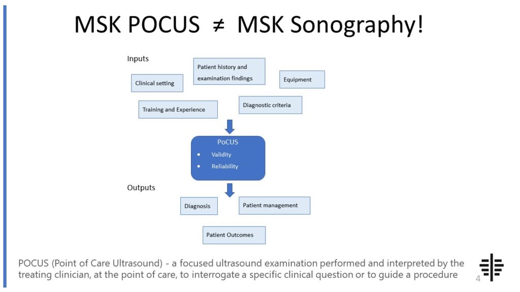
The POCUS process is very different to traditional US based in a radiology establishment. And POCUS practitioners need to be aware of those factors, unique to their particular situation, that influence diagnostic accuracy. That was the topic I presented at the plenary session of the NZAMM Annual Scientific Meeting in Wellington. A picture says 1000 ...

Posted by on 2022-10-04
We’re proud to announce that the New Zealand College of Musculoskeletal Medicine has endorsed our POCUS courses for CME and as part of vocational training. The NZCMM is responsible for setting the high standards and training of Specialist Musculoskeletal Medicine Physicians in New Zealand. NZCMM endorsement is an acknowledgement that our courses meet these standards. ...

Posted by on 2022-06-23
Ultrasound has several advantages for cartilage imaging. It is a non-invasive and cost-effective technique that does not involve exposure to ionizing radiation. Ultrasound provides real-time imaging, allowing for dynamic assessment of cartilage during joint movement. It can also be used to assess the thickness and integrity of the cartilage. However, ultrasound has limitations in terms of its ability to penetrate deep tissues and provide detailed images of the cartilage structure. It may not be as sensitive as other imaging techniques in detecting subtle cartilage abnormalities. Additionally, the operator's skill and experience can affect the quality and interpretation of ultrasound images.

Computed tomography (CT) scans can detect cartilage abnormalities, but they may not be as accurate as other imaging techniques. CT scans use X-rays to create cross-sectional images of the body, providing detailed information about the bone structure. While CT scans can indirectly visualize cartilage by assessing the joint space and detecting changes in bone structure, they may not provide as clear and detailed images of the cartilage itself compared to MRI or arthroscopy. CT scans are more commonly used to assess bone abnormalities or to guide surgical interventions rather than specifically evaluating cartilage health.
Specific markers or contrast agents can be used in cartilage imaging to enhance the visualization of cartilage. These agents can be injected into the joint to highlight the cartilage and improve the detection of abnormalities. One commonly used contrast agent is gadolinium, which is used in magnetic resonance imaging (MRI) to enhance the contrast between the cartilage and surrounding tissues. Other markers, such as dyes or nanoparticles, can also be used to target specific components of the cartilage, providing additional information about its composition and health.

Arthroscopy is a direct imaging technique that allows for the evaluation of cartilage damage. It involves inserting a small camera into the joint through a small incision, providing a real-time view of the cartilage surface. Arthroscopy allows for a detailed assessment of the cartilage, including the detection of surface irregularities, fissures, or areas of cartilage loss. It also allows for the evaluation of the surrounding structures, such as ligaments and tendons. However, arthroscopy is an invasive procedure that requires anesthesia and carries a risk of complications. It is typically reserved for cases where other imaging techniques have not provided sufficient information or when surgical intervention is planned.
There are several emerging technologies and advancements in cartilage imaging. One promising technique is optical coherence tomography (OCT), which uses light waves to create high-resolution images of tissues. OCT can provide detailed images of the cartilage structure and detect early signs of degeneration. Another emerging technology is magnetic resonance spectroscopy (MRS), which can assess the biochemical composition of cartilage and provide information about its health and integrity. Additionally, researchers are exploring the use of advanced imaging techniques, such as diffusion-weighted imaging and T1rho mapping, to further improve the detection and characterization of cartilage abnormalities. These advancements have the potential to enhance the diagnosis and management of cartilage conditions in the future.

Musculoskeletal ultrasound plays a crucial role in the evaluation of synovial sarcoma by providing valuable information about the tumor's location, size, and characteristics. This imaging technique utilizes high-frequency sound waves to create detailed images of the soft tissues and structures surrounding the affected area. By examining the synovial sarcoma with ultrasound, healthcare professionals can assess the tumor's extent of infiltration into adjacent tissues, identify any associated cystic or necrotic areas, and determine the presence of vascular involvement. Additionally, musculoskeletal ultrasound allows for real-time visualization, enabling the evaluation of dynamic changes in the tumor during movement or manipulation. This non-invasive and cost-effective imaging modality aids in the accurate diagnosis, staging, and monitoring of synovial sarcoma, ultimately guiding treatment decisions and improving patient outcomes.