
There are several types of joint imaging techniques used in medical diagnostics. One common technique is X-ray imaging, which uses electromagnetic radiation to produce images of bones and joints. X-rays can help identify fractures, dislocations, and bone abnormalities. Another technique is magnetic resonance imaging (MRI), which uses a strong magnetic field and radio waves to create detailed images of soft tissues, including joints. MRI is particularly useful for visualizing joint structures such as cartilage, ligaments, and tendons. Ultrasound imaging is also commonly used to assess joint inflammation and fluid accumulation. It uses high-frequency sound waves to create real-time images of the joint, allowing for the evaluation of joint fluid and the detection of abnormalities. Finally, computed tomography (CT) scans can provide detailed images of joint injuries and abnormalities by combining multiple X-ray images taken from different angles.
Magnetic resonance imaging (MRI) is a valuable tool for visualizing joint structures. It uses a strong magnetic field and radio waves to create detailed images of soft tissues, including joints. MRI can provide high-resolution images of the joint, allowing for the visualization of structures such as cartilage, ligaments, and tendons. It can help identify abnormalities such as tears, inflammation, and degenerative changes in these structures. MRI is particularly useful for diagnosing conditions such as osteoarthritis, rheumatoid arthritis, and meniscal tears. It can also help guide treatment decisions, such as determining the need for surgery or monitoring the effectiveness of treatment.
Over the last couple of years, we’ve brought you several courses focusing on Ultrasound Guided Injection Techniques. They’ve been extremely popular, and like our other courses, the feedback has been fantastic. One thing we’ve learnt along the way is that to get the most out of learning injection techniques, a solid grounding in MSK Ultrasound ...
Posted by on 2024-02-10
What a year 2023 was! We’ve loved bringing you courses covering US of the upper and lower limb, and US guided injections through the year. The mix of health professionals from all sorts of backgrounds (Doctors, Nurses, Physios, Sonographers to name a few) has been amazing to be part of. We’ve been humbled by your ...
Posted by on 2023-09-17
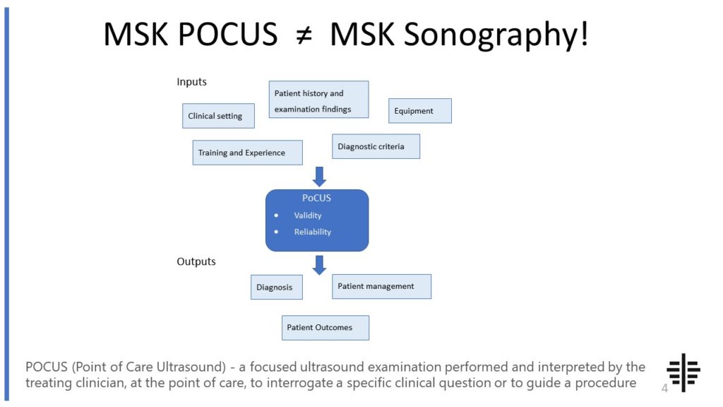
The POCUS process is very different to traditional US based in a radiology establishment. And POCUS practitioners need to be aware of those factors, unique to their particular situation, that influence diagnostic accuracy. That was the topic I presented at the plenary session of the NZAMM Annual Scientific Meeting in Wellington. A picture says 1000 ...

Posted by on 2022-10-04
We’re proud to announce that the New Zealand College of Musculoskeletal Medicine has endorsed our POCUS courses for CME and as part of vocational training. The NZCMM is responsible for setting the high standards and training of Specialist Musculoskeletal Medicine Physicians in New Zealand. NZCMM endorsement is an acknowledgement that our courses meet these standards. ...

Posted by on 2022-06-23
The RNZCUC has endorsed our courses as approved CME. We’re proud to be able to meet the training needs of Urgent Care Physicians, and look forward to meeting you at future courses.

Posted by on 2021-05-30
Ultrasound imaging plays a crucial role in assessing joint inflammation and fluid accumulation. It uses high-frequency sound waves to create real-time images of the joint, allowing for the evaluation of joint fluid and the detection of abnormalities. Ultrasound can help identify conditions such as synovitis, bursitis, and effusion, which are characterized by joint inflammation and fluid accumulation. It can also guide procedures such as joint aspirations or injections, providing real-time visualization of the needle placement. Ultrasound is a safe and non-invasive imaging technique that does not involve the use of ionizing radiation, making it suitable for repeated examinations and monitoring of joint conditions over time.

Computed tomography (CT) scans can provide detailed images of joint injuries and abnormalities. CT scans use X-rays and computer processing to create cross-sectional images of the body. They can provide high-resolution images of bones and joints, allowing for the detection of fractures, dislocations, and other bone abnormalities. CT scans are particularly useful for evaluating complex fractures, assessing joint alignment, and planning surgical interventions. However, CT scans involve exposure to ionizing radiation, which may limit their use in certain populations, such as pregnant women or children. Additionally, CT scans do not provide as much detail of soft tissues as other imaging techniques like MRI.
Arthrography is a technique that helps evaluate the integrity of joint cartilage and ligaments. It involves the injection of a contrast agent, such as a dye or a gas, into the joint space. The contrast agent helps outline the joint structures, making them more visible on X-ray or fluoroscopy images. Arthrography can help identify abnormalities such as tears in the joint cartilage or ligaments, as well as evaluate joint stability. It is commonly used in the diagnosis of conditions such as rotator cuff tears, labral tears, and ligament injuries. Arthrography can provide valuable information for treatment planning, such as determining the need for surgery or guiding minimally invasive interventions like joint injections.

Nuclear medicine imaging techniques have both advantages and limitations for evaluating joint disorders. One commonly used technique is bone scintigraphy, which involves the injection of a radioactive tracer that accumulates in areas of increased bone turnover or inflammation. Bone scintigraphy can help detect conditions such as osteoarthritis, infection, and bone tumors. It is particularly sensitive in detecting early changes in bone metabolism, even before structural abnormalities are visible on other imaging modalities. However, nuclear medicine imaging has limitations, including lower spatial resolution compared to techniques like MRI or CT scans. It also involves exposure to ionizing radiation, which may limit its use in certain populations.
Positron emission tomography (PET) imaging assists in detecting early signs of joint diseases like arthritis. PET imaging involves the injection of a radioactive tracer that is taken up by metabolically active tissues, such as areas of inflammation. PET scans can help identify areas of increased metabolic activity in the joints, which may indicate early signs of arthritis or other inflammatory conditions. PET imaging can provide valuable information for early diagnosis, monitoring disease progression, and assessing treatment response. However, PET imaging has limitations, including limited availability and higher cost compared to other imaging techniques. It is often used in combination with other imaging modalities to provide a comprehensive evaluation of joint diseases.

When performing musculoskeletal ultrasound on geriatric patients, there are several important considerations to keep in mind. Firstly, due to the natural aging process, geriatric patients may have decreased muscle mass and strength, as well as reduced joint mobility. This can affect the quality of the ultrasound images obtained and may require adjustments in the scanning technique. Additionally, geriatric patients may have underlying medical conditions such as arthritis or osteoporosis, which can affect the musculoskeletal structures being evaluated. It is crucial to take these conditions into account and tailor the ultrasound examination accordingly. Furthermore, the skin of geriatric patients may be more fragile and prone to injury, so care must be taken to ensure patient comfort and safety during the procedure. Lastly, communication and patient cooperation may be more challenging in geriatric patients, necessitating a calm and patient approach to obtain accurate and reliable ultrasound results.
Musculoskeletal ultrasound can be a useful tool for diagnosing sacral stress fractures. This imaging technique utilizes high-frequency sound waves to create detailed images of the musculoskeletal system, allowing for the visualization of bone structures and potential fractures. By examining the sacrum using musculoskeletal ultrasound, healthcare professionals can identify signs of stress fractures, such as cortical irregularities, periosteal reactions, and bone edema. Additionally, musculoskeletal ultrasound can provide real-time imaging, allowing for dynamic assessment of the sacrum during movement or stress tests. This can aid in the accurate diagnosis of sacral stress fractures and help guide appropriate treatment plans.
Musculoskeletal ultrasound plays a crucial role in diagnosing intra-articular pathology by providing detailed imaging of the joint structures and surrounding soft tissues. This imaging technique utilizes high-frequency sound waves to create real-time images, allowing for the visualization of intra-articular structures such as the synovium, articular cartilage, ligaments, and tendons. By assessing the integrity and morphology of these structures, musculoskeletal ultrasound can help identify various pathologies, including synovitis, joint effusion, cartilage defects, ligament tears, and tendon abnormalities. Additionally, musculoskeletal ultrasound enables the evaluation of joint movement and function, aiding in the assessment of dynamic intra-articular conditions. The use of musculoskeletal ultrasound in diagnosing intra-articular pathology offers a non-invasive and cost-effective imaging modality that can provide valuable information for treatment planning and monitoring the response to therapy.
Ultrasound offers several advantages over MRI for diagnosing musculoskeletal disorders. Firstly, ultrasound is a non-invasive and painless imaging technique that does not involve exposure to ionizing radiation, making it a safer option for patients, especially those who may require multiple imaging sessions. Additionally, ultrasound provides real-time imaging, allowing for dynamic assessment of the musculoskeletal system during movement or stress tests. This real-time capability enables the visualization of soft tissues, such as tendons, ligaments, and muscles, in their natural state, providing valuable information about their structure and function. Moreover, ultrasound is more cost-effective and readily available compared to MRI, making it a more accessible diagnostic tool for musculoskeletal disorders. Overall, the use of ultrasound in diagnosing musculoskeletal disorders offers numerous benefits, including safety, real-time imaging, and cost-effectiveness.