
Ultrasonography is a medical imaging technique that uses high-frequency sound waves to create real-time images of various structures within the body. It is particularly useful for visualizing key structures such as the liver, gallbladder, kidneys, bladder, uterus, ovaries, prostate, and blood vessels. These structures can be visualized in detail, allowing healthcare professionals to assess their size, shape, and overall condition. Ultrasonography can also be used to guide procedures such as biopsies or needle aspirations, providing real-time visualization and increased accuracy.
Ultrasonography differentiates between different types of tissues based on their acoustic properties. When sound waves pass through different tissues, they are reflected back at different rates and intensities. This information is then processed by the ultrasound machine to create an image. For example, dense tissues like bone or calcifications reflect sound waves strongly, appearing bright on the ultrasound image. On the other hand, fluid-filled structures like cysts or blood vessels allow sound waves to pass through easily, appearing dark on the image. By analyzing these differences in acoustic properties, healthcare professionals can distinguish between different types of tissues and identify any abnormalities.
Over the last couple of years, we’ve brought you several courses focusing on Ultrasound Guided Injection Techniques. They’ve been extremely popular, and like our other courses, the feedback has been fantastic. One thing we’ve learnt along the way is that to get the most out of learning injection techniques, a solid grounding in MSK Ultrasound ...
Posted by on 2024-02-10
What a year 2023 was! We’ve loved bringing you courses covering US of the upper and lower limb, and US guided injections through the year. The mix of health professionals from all sorts of backgrounds (Doctors, Nurses, Physios, Sonographers to name a few) has been amazing to be part of. We’ve been humbled by your ...
Posted by on 2023-09-17
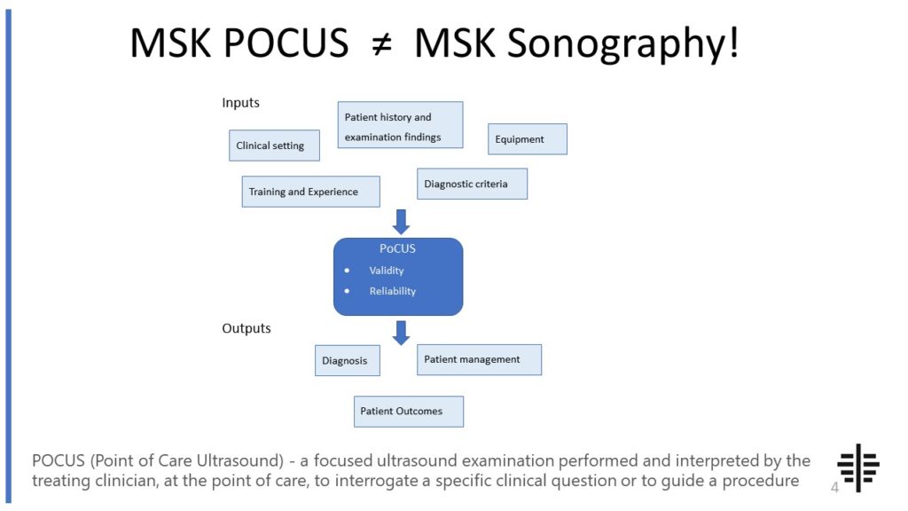
The POCUS process is very different to traditional US based in a radiology establishment. And POCUS practitioners need to be aware of those factors, unique to their particular situation, that influence diagnostic accuracy. That was the topic I presented at the plenary session of the NZAMM Annual Scientific Meeting in Wellington. A picture says 1000 ...

Posted by on 2022-10-04
We’re proud to announce that the New Zealand College of Musculoskeletal Medicine has endorsed our POCUS courses for CME and as part of vocational training. The NZCMM is responsible for setting the high standards and training of Specialist Musculoskeletal Medicine Physicians in New Zealand. NZCMM endorsement is an acknowledgement that our courses meet these standards. ...

Posted by on 2022-06-23
There are several advantages of using ultrasonography over other imaging techniques. Firstly, it is a non-invasive procedure that does not involve the use of ionizing radiation, making it safe for both patients and healthcare professionals. Secondly, ultrasonography provides real-time imaging, allowing for dynamic assessment of structures and functions. This is particularly useful in procedures such as guiding needle placements or monitoring blood flow. Additionally, ultrasonography is relatively inexpensive compared to other imaging modalities, making it more accessible in various healthcare settings. It is also portable and can be performed at the bedside, making it convenient for point-of-care assessments.

The Doppler effect plays a crucial role in assessing blood flow using ultrasonography. The Doppler effect refers to the change in frequency of sound waves when there is relative motion between the source of the sound waves and the observer. In the context of ultrasonography, this effect is used to assess the direction and speed of blood flow within blood vessels. By emitting sound waves at a specific frequency and analyzing the frequency shift of the reflected waves, healthcare professionals can determine the velocity and direction of blood flow. This information is then displayed as color-coded images or waveforms, providing valuable insights into the vascular system and helping diagnose conditions such as deep vein thrombosis or arterial stenosis.
One limitation of ultrasonography is its limited ability to visualize deep structures within the body. Sound waves used in ultrasonography have difficulty penetrating through dense tissues such as bone or air-filled structures like the lungs. This can make it challenging to visualize structures located deep within the body, especially in obese patients or in cases where there are significant barriers to sound wave transmission. In such situations, other imaging modalities like computed tomography (CT) or magnetic resonance imaging (MRI) may be more suitable for obtaining detailed images of deep structures.

The operator's skill and experience play a significant role in the quality of ultrasonographic images. The operator needs to have a thorough understanding of anatomy, physiology, and the principles of ultrasonography to obtain accurate and meaningful images. They must be able to adjust various settings on the ultrasound machine, such as the frequency and gain, to optimize image quality. Additionally, the operator's ability to position the transducer correctly and interpret the images in real-time is crucial. Experience and proficiency in ultrasonography can greatly enhance the diagnostic accuracy and efficiency of the procedure.
Ultrasonography has a wide range of applications in medical diagnosis and monitoring. It is commonly used in obstetrics to monitor fetal development, assess the placenta, and detect any abnormalities. In cardiology, ultrasonography helps evaluate the structure and function of the heart, including the valves, chambers, and blood flow patterns. It is also used in the diagnosis and monitoring of various conditions such as gallstones, kidney stones, liver diseases, thyroid nodules, and musculoskeletal disorders. Additionally, ultrasonography is frequently used in interventional procedures, such as guiding the placement of central venous catheters or performing ultrasound-guided biopsies. Its versatility, safety, and real-time imaging capabilities make it an invaluable tool in various medical specialties.

In musculoskeletal ultrasound of patients with rheumatoid arthritis, typical findings include synovial hypertrophy, joint effusion, and power Doppler signal. Synovial hypertrophy refers to the thickening of the synovial lining of the joints, which is a characteristic feature of rheumatoid arthritis. Joint effusion, or the accumulation of fluid within the joint space, is commonly observed in patients with this condition. Power Doppler signal, which detects blood flow within the synovium, is often increased in rheumatoid arthritis due to the inflammation and increased vascularity associated with the disease. Other findings may include erosions, tenosynovitis, and bursitis, which are indicative of the destructive nature of rheumatoid arthritis on the musculoskeletal system. Overall, musculoskeletal ultrasound plays a valuable role in the assessment and monitoring of rheumatoid arthritis, providing important information about disease activity and progression.
Osteitis pubis is a condition characterized by inflammation of the pubic symphysis and surrounding structures. When evaluating patients with osteitis pubis using musculoskeletal ultrasound, typical findings may include thickening and irregularity of the pubic symphysis, increased vascularity in the affected area, and the presence of fluid collections or edema. Additionally, ultrasound may reveal tendon abnormalities, such as tendonitis or tears, in the adjacent muscles, such as the adductor muscles or rectus abdominis. The ultrasound may also show signs of bursitis or inflammation of the bursae surrounding the pubic symphysis. These findings are important in diagnosing and monitoring the progression of osteitis pubis, as well as guiding treatment decisions.
Musculoskeletal ultrasound plays a crucial role in the evaluation of soft tissue masses by providing detailed imaging of the affected area. This imaging technique utilizes high-frequency sound waves to create real-time images of the soft tissues, allowing for the identification and characterization of masses. By visualizing the size, shape, location, and internal structure of the mass, musculoskeletal ultrasound aids in determining the nature of the lesion, such as whether it is a benign or malignant tumor, a cyst, or an abscess. Additionally, musculoskeletal ultrasound can assess the vascularity of the mass by utilizing Doppler imaging, which helps in differentiating between solid and cystic lesions. Furthermore, this imaging modality allows for dynamic evaluation, enabling the assessment of the mass during movement or stress maneuvers, which can provide valuable information about its behavior and potential impact on nearby structures. Overall, musculoskeletal ultrasound is an invaluable tool in the assessment of soft tissue masses, aiding in accurate diagnosis and guiding appropriate management decisions.
Musculoskeletal ultrasound has proven to be a valuable tool in identifying synovitis in patients with inflammatory arthritis. This imaging technique utilizes high-frequency sound waves to produce detailed images of the musculoskeletal system, allowing for the visualization of synovial inflammation. By assessing the synovial membrane and joint space, musculoskeletal ultrasound can detect signs of synovitis, such as synovial thickening, increased vascularity, and effusion. Additionally, this modality enables the assessment of other related features, including erosions, tenosynovitis, and bursitis, providing a comprehensive evaluation of the inflammatory process. The use of musculoskeletal ultrasound in the diagnosis and monitoring of synovitis in patients with inflammatory arthritis has shown promising results, offering a non-invasive and cost-effective alternative to other imaging modalities.
Performing musculoskeletal ultrasound on obese patients presents several technical challenges. One of the main challenges is the increased depth of penetration required to visualize the musculoskeletal structures due to the thicker subcutaneous fat layer. This can result in reduced image quality and difficulty in identifying anatomical landmarks. Additionally, the increased attenuation of sound waves in obese patients can lead to decreased signal strength and decreased resolution of the ultrasound images. The larger body habitus of obese patients can also make it more challenging to position the ultrasound probe correctly and maintain adequate contact with the skin. Furthermore, the increased adipose tissue can cause acoustic shadowing, making it difficult to visualize structures that lie deep to the fat layer. These technical challenges highlight the importance of using appropriate ultrasound settings, selecting the appropriate transducer, and employing proper scanning techniques to optimize image quality and diagnostic accuracy in musculoskeletal ultrasound examinations of obese patients.