
There are several different types of ultrasound imaging techniques that are used in medical diagnosis. One common technique is 2D ultrasound, which uses high-frequency sound waves to create a two-dimensional image of the body. Another technique is 3D ultrasound, which uses multiple 2D images to create a three-dimensional image. Doppler ultrasound is used to assess blood flow and can be used to detect conditions such as deep vein thrombosis or evaluate the blood flow in the heart. Additionally, there is also contrast-enhanced ultrasound, which involves the use of contrast agents to enhance the visibility of certain structures or blood vessels.
Ultrasound works by emitting high-frequency sound waves into the body and then detecting the echoes that bounce back. A transducer is used to both emit the sound waves and receive the echoes. The sound waves travel through the body and when they encounter different tissues or structures, some of the sound waves are reflected back to the transducer. The transducer then converts these echoes into electrical signals, which are processed by a computer to create an image. The image is displayed on a monitor and can be interpreted by a healthcare professional.
Over the last couple of years, we’ve brought you several courses focusing on Ultrasound Guided Injection Techniques. They’ve been extremely popular, and like our other courses, the feedback has been fantastic. One thing we’ve learnt along the way is that to get the most out of learning injection techniques, a solid grounding in MSK Ultrasound ...
Posted by on 2024-02-10
What a year 2023 was! We’ve loved bringing you courses covering US of the upper and lower limb, and US guided injections through the year. The mix of health professionals from all sorts of backgrounds (Doctors, Nurses, Physios, Sonographers to name a few) has been amazing to be part of. We’ve been humbled by your ...
Posted by on 2023-09-17
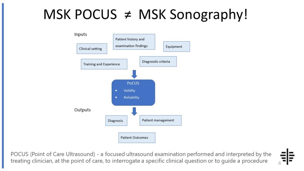
The POCUS process is very different to traditional US based in a radiology establishment. And POCUS practitioners need to be aware of those factors, unique to their particular situation, that influence diagnostic accuracy. That was the topic I presented at the plenary session of the NZAMM Annual Scientific Meeting in Wellington. A picture says 1000 ...

Posted by on 2022-10-04
We’re proud to announce that the New Zealand College of Musculoskeletal Medicine has endorsed our POCUS courses for CME and as part of vocational training. The NZCMM is responsible for setting the high standards and training of Specialist Musculoskeletal Medicine Physicians in New Zealand. NZCMM endorsement is an acknowledgement that our courses meet these standards. ...

Posted by on 2022-06-23
There are several advantages of using ultrasound over other imaging modalities. One major advantage is that ultrasound does not involve the use of ionizing radiation, making it a safer option for both patients and healthcare professionals. Ultrasound is also non-invasive and painless, making it well-tolerated by patients. It can be performed quickly and in real-time, allowing for immediate assessment and diagnosis. Additionally, ultrasound is relatively inexpensive compared to other imaging modalities and can be easily portable, making it accessible in various healthcare settings.

Despite its many advantages, ultrasound imaging does have some limitations. One limitation is that ultrasound is highly operator-dependent, meaning that the quality of the images can vary depending on the skill and experience of the person performing the examination. Additionally, ultrasound is not able to penetrate bone or air, so it may not be suitable for imaging certain areas of the body. The image quality can also be affected by factors such as obesity or the presence of gas in the intestines. Finally, ultrasound may not be able to provide as detailed anatomical information as other imaging modalities such as CT or MRI.
Ultrasound has a wide range of applications in medical diagnosis. It is commonly used to evaluate the abdomen, including the liver, gallbladder, kidneys, and pancreas. It can also be used to assess the thyroid gland, breasts, and musculoskeletal system. In addition, ultrasound is frequently used in obstetrics to monitor the development of the fetus and assess the health of the mother during pregnancy. It is also used in cardiology to evaluate the structure and function of the heart. Ultrasound-guided procedures, such as biopsies or fluid drainage, are also common.

In obstetrics and gynecology, ultrasound plays a crucial role in the assessment of pregnancy and reproductive health. It is used to confirm pregnancy, determine the gestational age, and monitor the growth and development of the fetus. Ultrasound can also detect abnormalities in the uterus, ovaries, and fallopian tubes, such as fibroids or ovarian cysts. It is used to guide procedures such as amniocentesis or fetal blood sampling. In gynecology, ultrasound is used to evaluate conditions such as endometriosis, polycystic ovary syndrome, or uterine abnormalities.
When performing ultrasound examinations, certain safety precautions should be taken. It is important to use the lowest possible power settings to minimize the exposure of the patient to sound waves. The transducer should be properly disinfected before and after each use to prevent the spread of infection. The examination room should be clean and well-maintained, and the healthcare professional should follow proper hygiene practices, such as wearing gloves and using sterile gel. It is also important to obtain informed consent from the patient and ensure their comfort and privacy throughout the examination.

Typical findings in musculoskeletal ultrasound of patients with tendon ruptures include discontinuity or complete absence of the tendon fibers, focal hypoechoic or anechoic areas representing the gap in the tendon, and retraction of the torn ends. Additionally, there may be surrounding edema, hematoma, or fluid accumulation in the tendon sheath. The ultrasound may also reveal thickening or irregularity of the tendon edges, indicating chronic degenerative changes. Doppler imaging can be used to assess vascularity and rule out associated vascular injury. Overall, musculoskeletal ultrasound plays a crucial role in the diagnosis and evaluation of tendon ruptures, providing valuable information for treatment planning and monitoring the healing process.
Musculoskeletal ultrasound has been found to be an effective diagnostic tool for meniscal tears. This imaging technique utilizes high-frequency sound waves to produce detailed images of the musculoskeletal system, including the knee joint. By visualizing the meniscus, which is a cartilage structure in the knee, ultrasound can help identify tears or other abnormalities. The use of musculoskeletal ultrasound in diagnosing meniscal tears offers several advantages, such as its non-invasive nature, real-time imaging capabilities, and the ability to assess both the structure and function of the meniscus. Additionally, ultrasound can be performed at the point of care, making it a convenient and accessible option for patients. Overall, musculoskeletal ultrasound is a valuable tool in the diagnosis of meniscal tears, providing accurate and timely information for appropriate management and treatment decisions.
Musculoskeletal ultrasound is a valuable tool for assessing spinal pathology, but it does have some limitations. One limitation is that it may not provide a comprehensive view of the entire spine. Due to the limited field of view, it may be challenging to visualize structures that are deep within the spine or located in areas that are difficult to access. Additionally, musculoskeletal ultrasound may not be as effective in evaluating bony structures, such as the vertebrae, as it is primarily designed to assess soft tissues. This means that it may not be able to detect certain types of spinal pathology, such as fractures or tumors. Furthermore, the quality of the ultrasound images can be affected by factors such as patient body habitus, operator skill, and patient cooperation, which may limit its accuracy and reliability in some cases. Therefore, while musculoskeletal ultrasound can be a useful tool for assessing spinal pathology, it should be used in conjunction with other imaging modalities, such as MRI or CT, to ensure a comprehensive evaluation.
Septic arthritis is an inflammatory condition of the joints caused by an infection. When examining the affected joint using sonography, several characteristic features can be observed. These include the presence of joint effusion, which is an accumulation of fluid within the joint space. The effusion may appear hypoechoic or anechoic on the ultrasound image. In addition, there may be synovial thickening, which is an increase in the thickness of the synovial lining of the joint. This can be visualized as a hypoechoic or hyperechoic area surrounding the joint. Another sonographic feature of septic arthritis is the presence of synovial debris, which can appear as echogenic material within the joint space. Doppler imaging may also reveal increased vascularity within the synovium, indicating an inflammatory response. Overall, sonography can be a valuable tool in the diagnosis of septic arthritis, allowing for the visualization of these characteristic features and guiding appropriate treatment.
Musculoskeletal ultrasound is a valuable imaging modality that can aid in the differentiation between benign and malignant bone tumors. By utilizing high-frequency sound waves, musculoskeletal ultrasound can provide detailed images of the bone and surrounding soft tissues, allowing for the assessment of various characteristics of the tumor. These characteristics include size, shape, vascularity, and internal architecture. Additionally, musculoskeletal ultrasound can help identify specific features such as cortical disruption, periosteal reaction, and invasion of adjacent structures, which are indicative of malignancy. Furthermore, the use of Doppler ultrasound can assess the blood flow within the tumor, providing additional information for differentiation. While musculoskeletal ultrasound is a valuable tool, it is important to note that it may not be able to definitively differentiate between all benign and malignant bone tumors. In such cases, further imaging modalities, such as magnetic resonance imaging (MRI) or biopsy, may be necessary for a more accurate diagnosis.