
There are several imaging techniques used to visualize ligaments. One common technique is magnetic resonance imaging (MRI), which uses a strong magnetic field and radio waves to create detailed images of the ligaments. Another technique is ultrasound, which uses high-frequency sound waves to produce real-time images of the ligaments. Computed tomography (CT) scans can also be used to evaluate ligament damage by taking multiple X-ray images from different angles and creating a cross-sectional view. Additionally, arthrography involves injecting a contrast dye into the joint and then taking X-ray images to visualize the ligaments.
Magnetic resonance imaging (MRI) is a valuable tool in the diagnosis of ligament injuries. It provides detailed images of the ligaments, allowing healthcare professionals to assess the extent of the injury and determine the appropriate treatment plan. MRI can detect ligament tears, sprains, and other abnormalities with high accuracy. It also allows for the visualization of surrounding structures, such as bones and soft tissues, which can help in identifying any additional damage. MRI is non-invasive and does not involve exposure to ionizing radiation, making it a safe option for imaging ligament injuries.
Over the last couple of years, we’ve brought you several courses focusing on Ultrasound Guided Injection Techniques. They’ve been extremely popular, and like our other courses, the feedback has been fantastic. One thing we’ve learnt along the way is that to get the most out of learning injection techniques, a solid grounding in MSK Ultrasound ...
Posted by on 2024-02-10
What a year 2023 was! We’ve loved bringing you courses covering US of the upper and lower limb, and US guided injections through the year. The mix of health professionals from all sorts of backgrounds (Doctors, Nurses, Physios, Sonographers to name a few) has been amazing to be part of. We’ve been humbled by your ...
Posted by on 2023-09-17
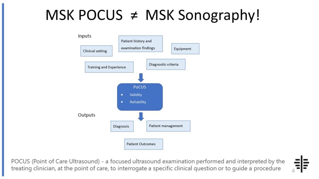
The POCUS process is very different to traditional US based in a radiology establishment. And POCUS practitioners need to be aware of those factors, unique to their particular situation, that influence diagnostic accuracy. That was the topic I presented at the plenary session of the NZAMM Annual Scientific Meeting in Wellington. A picture says 1000 ...

Posted by on 2022-10-04
We’re proud to announce that the New Zealand College of Musculoskeletal Medicine has endorsed our POCUS courses for CME and as part of vocational training. The NZCMM is responsible for setting the high standards and training of Specialist Musculoskeletal Medicine Physicians in New Zealand. NZCMM endorsement is an acknowledgement that our courses meet these standards. ...

Posted by on 2022-06-23
The RNZCUC has endorsed our courses as approved CME. We’re proud to be able to meet the training needs of Urgent Care Physicians, and look forward to meeting you at future courses.

Posted by on 2021-05-30
Ultrasound has both advantages and disadvantages when it comes to imaging ligaments. One advantage is that it is a real-time imaging technique, meaning that it can show the movement and function of the ligaments. It is also non-invasive and does not involve exposure to ionizing radiation. However, ultrasound has limitations in terms of its ability to visualize deep structures and provide detailed images. It may not be as effective in detecting subtle ligament injuries compared to other imaging techniques like MRI. Additionally, the quality of the ultrasound images can be operator-dependent, meaning that the skill and experience of the technician performing the ultrasound can affect the accuracy of the results.

X-rays are not typically used to detect ligament injuries directly. X-rays are best suited for visualizing bones and can help identify fractures or dislocations that may be associated with ligament injuries. However, ligaments themselves do not show up well on X-ray images. In some cases, X-rays may show indirect signs of ligament injuries, such as joint instability or abnormal alignment. If a healthcare professional suspects a ligament injury based on the patient's symptoms and physical examination, they may order additional imaging tests like MRI or ultrasound for a more accurate diagnosis.
Computed tomography (CT) scans can play a role in evaluating ligament damage, particularly in cases where a more detailed assessment is needed. CT scans use X-rays and computer processing to create cross-sectional images of the ligaments and surrounding structures. This can help in identifying fractures, dislocations, or other bony abnormalities that may be associated with ligament injuries. CT scans can provide a more detailed view of the ligaments compared to X-rays alone. However, like X-rays, CT scans do involve exposure to ionizing radiation, which should be considered when determining the appropriate imaging modality for ligament evaluation.

In sports medicine, there are specific ligament imaging techniques that are commonly used. One such technique is stress radiography, which involves applying stress to the joint while taking X-ray images. This can help assess the stability of the ligaments and detect any abnormal movement or laxity. Another technique is dynamic ultrasound, which involves performing ultrasound imaging while the joint is in motion. This can provide valuable information about the function and integrity of the ligaments during activity. Sports medicine professionals may also use arthroscopy, a minimally invasive procedure that involves inserting a small camera into the joint to directly visualize the ligaments and other structures.
Arthrography is a technique that assists in the visualization of ligaments by injecting a contrast dye into the joint and then taking X-ray images. The contrast dye helps to highlight the ligaments and other structures within the joint, making them more visible on the X-ray images. Arthrography can provide detailed information about the integrity and function of the ligaments, as well as any abnormalities or injuries. It is particularly useful in evaluating ligament tears, joint instability, and other conditions that may require surgical intervention. Arthrography is a minimally invasive procedure that can be performed on an outpatient basis, making it a convenient option for ligament imaging.

Musculoskeletal ultrasound has shown promise in the diagnosis of pigmented villonodular synovitis (PVNS). Several studies have demonstrated the effectiveness of ultrasound in detecting the characteristic features of PVNS, such as synovial thickening, joint effusion, and the presence of nodules or villi. The use of high-frequency transducers and Doppler imaging can provide additional information about the vascularity of the synovial tissue, which is often increased in PVNS. However, it is important to note that ultrasound findings should be correlated with clinical and histopathological findings for a definitive diagnosis of PVNS. Other imaging modalities, such as magnetic resonance imaging (MRI), may also be used in conjunction with ultrasound to improve diagnostic accuracy. Overall, musculoskeletal ultrasound can be a valuable tool in the diagnosis of PVNS, but it should be used in combination with other diagnostic methods for a comprehensive evaluation.
Musculoskeletal ultrasound plays a crucial role in the diagnosis of peripheral nerve tumors by providing detailed imaging of the affected area. This imaging technique utilizes high-frequency sound waves to create real-time images of the musculoskeletal system, allowing for the visualization of nerve structures and any abnormalities present. By using musculoskeletal ultrasound, healthcare professionals can accurately identify the location, size, and characteristics of peripheral nerve tumors, such as schwannomas or neurofibromas. Additionally, this imaging modality enables the assessment of surrounding tissues, including muscles, tendons, and ligaments, which can help determine the extent of tumor involvement and potential compression of adjacent structures. Overall, musculoskeletal ultrasound aids in the early detection and precise localization of peripheral nerve tumors, facilitating timely and appropriate management strategies.
Musculoskeletal ultrasound is a valuable imaging technique that can aid in the differentiation of various types of muscle tumors. This non-invasive procedure utilizes high-frequency sound waves to produce detailed images of the musculoskeletal system, allowing for the visualization of soft tissues, muscles, and tumors. By assessing the size, shape, location, and characteristics of the tumor, musculoskeletal ultrasound can help distinguish between different types of muscle tumors, such as rhabdomyosarcoma, leiomyosarcoma, and liposarcoma. Additionally, this imaging modality can provide information about the vascularity of the tumor, which can further aid in the diagnosis and classification of the tumor. Overall, musculoskeletal ultrasound plays a crucial role in the evaluation and management of muscle tumors, providing valuable insights for accurate diagnosis and appropriate treatment planning.
Musculoskeletal ultrasound is a valuable tool for assessing joint effusions, but it does have some limitations. One limitation is that it may not be able to accurately detect small or subtle effusions, especially in deep joints or joints with complex anatomy. Additionally, the operator's skill and experience can greatly impact the accuracy of the ultrasound findings. In some cases, the presence of gas or air in the joint can also hinder the visualization of the effusion. Furthermore, ultrasound may not be able to differentiate between different types of joint effusions, such as inflammatory or infectious effusions, which may require additional diagnostic tests. Overall, while musculoskeletal ultrasound is a useful imaging modality for assessing joint effusions, it is important to consider its limitations and use it in conjunction with other diagnostic tools for a comprehensive evaluation.
Musculoskeletal ultrasound plays a crucial role in the diagnosis of tendonitis by providing detailed imaging of the affected tendons. This imaging technique utilizes high-frequency sound waves to create real-time images of the musculoskeletal system, allowing healthcare professionals to visualize the tendon structure and identify any abnormalities or inflammation. By using musculoskeletal ultrasound, doctors can accurately assess the thickness, integrity, and vascularity of the tendons, which are key indicators of tendonitis. Additionally, this imaging modality enables the evaluation of surrounding structures such as muscles, ligaments, and bursae, providing a comprehensive assessment of the affected area. The ability to visualize the tendon in real-time and assess its dynamic function during movement further aids in the diagnosis and management of tendonitis. Overall, musculoskeletal ultrasound is a valuable tool that enhances the diagnostic accuracy and guides appropriate treatment strategies for tendonitis.
Musculoskeletal ultrasound has been found to be highly effective in diagnosing rotator cuff injuries. This imaging technique utilizes sound waves to create detailed images of the musculoskeletal structures, allowing for the visualization of the rotator cuff tendons and surrounding tissues. By assessing the thickness, integrity, and any abnormalities in the rotator cuff tendons, musculoskeletal ultrasound can accurately identify rotator cuff tears, tendinitis, and other related injuries. Additionally, this diagnostic tool enables the evaluation of the subacromial space, bursa, and other structures involved in rotator cuff pathology. The real-time nature of musculoskeletal ultrasound also allows for dynamic assessment of the rotator cuff during movement, providing valuable information about impingement and muscle function. Overall, musculoskeletal ultrasound is a valuable and reliable tool for diagnosing rotator cuff injuries, offering clinicians a non-invasive and cost-effective imaging modality.