
Ultrasound can be used to diagnose metabolic diseases affecting the musculoskeletal system by providing real-time imaging of the affected area. It can help visualize changes in the soft tissues, such as muscle and tendon abnormalities, as well as detect fluid accumulation or inflammation. Ultrasound can also assess blood flow to the area, which can be useful in evaluating conditions such as vasculitis or ischemia. Additionally, ultrasound can be used to guide needle placement for diagnostic or therapeutic procedures, such as joint aspirations or injections, providing further information about the underlying metabolic disease.
Ultrasound for Neurological Disorders Affecting the Musculoskeletal System
There are several advantages of using ultrasound over other imaging techniques for detecting metabolic diseases in the musculoskeletal system. Firstly, ultrasound is non-invasive and does not involve exposure to ionizing radiation, making it a safer option, especially for repeated or long-term monitoring. Secondly, ultrasound is readily available, portable, and relatively inexpensive compared to other imaging modalities, making it more accessible for both patients and healthcare providers. Additionally, ultrasound provides real-time imaging, allowing for dynamic assessment of the affected area, which can be particularly useful in evaluating functional abnormalities or changes over time.
Over the last couple of years, we’ve brought you several courses focusing on Ultrasound Guided Injection Techniques. They’ve been extremely popular, and like our other courses, the feedback has been fantastic. One thing we’ve learnt along the way is that to get the most out of learning injection techniques, a solid grounding in MSK Ultrasound ...
Posted by on 2024-02-10
What a year 2023 was! We’ve loved bringing you courses covering US of the upper and lower limb, and US guided injections through the year. The mix of health professionals from all sorts of backgrounds (Doctors, Nurses, Physios, Sonographers to name a few) has been amazing to be part of. We’ve been humbled by your ...
Posted by on 2023-09-17
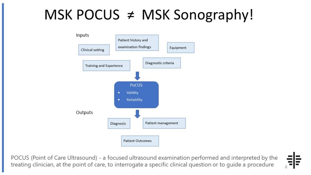
The POCUS process is very different to traditional US based in a radiology establishment. And POCUS practitioners need to be aware of those factors, unique to their particular situation, that influence diagnostic accuracy. That was the topic I presented at the plenary session of the NZAMM Annual Scientific Meeting in Wellington. A picture says 1000 ...

Posted by on 2022-10-04
We’re proud to announce that the New Zealand College of Musculoskeletal Medicine has endorsed our POCUS courses for CME and as part of vocational training. The NZCMM is responsible for setting the high standards and training of Specialist Musculoskeletal Medicine Physicians in New Zealand. NZCMM endorsement is an acknowledgement that our courses meet these standards. ...

Posted by on 2022-06-23
Ultrasound can accurately differentiate between different types of metabolic diseases affecting the musculoskeletal system to some extent. However, the specific diagnosis may require additional imaging or laboratory tests for confirmation. Ultrasound can provide valuable information about the structural changes in the affected tissues, such as muscle atrophy, tendon thickening, or joint effusion. It can also help identify specific patterns or characteristics associated with certain metabolic diseases, such as the presence of tophi in gout or synovial hypertrophy in rheumatoid arthritis. However, a comprehensive evaluation often involves a combination of clinical assessment, imaging, and laboratory investigations.

While ultrasound is a valuable tool for diagnosing metabolic diseases in the musculoskeletal system, it does have some limitations and drawbacks. One limitation is the operator-dependency, as the quality of the ultrasound images can vary depending on the skill and experience of the sonographer. Additionally, ultrasound may not be able to visualize deeper structures or areas that are difficult to access, such as the spine or deep joints. In some cases, ultrasound may also have limited sensitivity in detecting certain metabolic diseases, especially in the early stages or when the changes are subtle. Therefore, it is important to consider the clinical context and potentially use other imaging modalities or tests to complement the ultrasound findings.
Ultrasound helps in monitoring the progression or treatment of metabolic diseases affecting the musculoskeletal system by providing serial imaging of the affected area. It can help assess the response to treatment, such as the reduction in inflammation or the healing of injured tissues. Ultrasound can also be used to guide interventions, such as needle aspirations or injections, to monitor the effectiveness of the procedure. Additionally, ultrasound can provide real-time imaging during functional assessments, such as evaluating muscle strength or joint mobility, which can be useful in tracking the functional improvement or deterioration over time.

There are several specific ultrasound techniques or protocols commonly used for evaluating metabolic diseases in the musculoskeletal system. One commonly used technique is grayscale ultrasound, which provides detailed anatomical information about the affected area. Doppler ultrasound, on the other hand, can assess blood flow and detect abnormalities such as vasculitis or ischemia. Power Doppler ultrasound is particularly useful in detecting low-flow or slow-flow conditions. Additionally, ultrasound elastography can assess tissue stiffness or elasticity, which can be altered in certain metabolic diseases. These different techniques can be used in combination to provide a comprehensive evaluation of the musculoskeletal system.
Ultrasound can detect various findings or abnormalities in patients with metabolic diseases affecting the musculoskeletal system. These findings may include muscle atrophy, tendon thickening or tears, joint effusion, synovial hypertrophy, or the presence of tophi. Ultrasound can also help identify signs of inflammation, such as increased vascularity or synovial thickening. Additionally, ultrasound can detect changes in the bone, such as erosions or cysts, which may be indicative of underlying metabolic diseases. By visualizing these abnormalities, ultrasound can aid in the diagnosis, monitoring, and management of metabolic diseases affecting the musculoskeletal system.

Musculoskeletal ultrasound plays a crucial role in diagnosing stress fractures in the foot by providing detailed imaging of the affected area. This non-invasive imaging technique utilizes high-frequency sound waves to create real-time images of the musculoskeletal structures, including bones, tendons, and ligaments. By using musculoskeletal ultrasound, healthcare professionals can visualize the specific location and extent of the stress fracture, allowing for accurate diagnosis and appropriate treatment planning. Additionally, this imaging modality can help differentiate stress fractures from other foot conditions, such as tendonitis or ligament sprains, by assessing the integrity of the surrounding soft tissues. The ability to visualize the fracture site in real-time and from multiple angles enhances the diagnostic accuracy and aids in monitoring the healing progress of the stress fracture. Overall, musculoskeletal ultrasound is a valuable tool in the diagnosis and management of stress fractures in the foot, providing clinicians with detailed and reliable information for optimal patient care.
Musculoskeletal ultrasound has the potential to differentiate between different types of soft tissue tumors. This imaging technique utilizes sound waves to create detailed images of the musculoskeletal system, allowing for the visualization of various soft tissue structures. By analyzing the characteristics of the tumor, such as its size, shape, vascularity, and echogenicity, musculoskeletal ultrasound can provide valuable information that can aid in the differentiation of different types of soft tissue tumors. Additionally, the use of Doppler ultrasound can assess the blood flow within the tumor, which can further contribute to the identification and classification of the tumor. However, it is important to note that while musculoskeletal ultrasound can provide valuable insights, it may not always be able to definitively differentiate between all types of soft tissue tumors. In such cases, additional imaging modalities or biopsy may be necessary for a more accurate diagnosis.
Musculoskeletal ultrasound can be a useful tool in diagnosing myositis. This imaging technique allows for the visualization of the muscles and surrounding tissues, providing valuable information about the presence of inflammation, muscle fiber changes, and other abnormalities associated with myositis. By using high-frequency sound waves, musculoskeletal ultrasound can detect muscle edema, muscle thickening, and the presence of muscle nodules, which are characteristic features of myositis. Additionally, this imaging modality can help differentiate between different types of myositis, such as polymyositis and dermatomyositis, by assessing the pattern and distribution of muscle involvement. Overall, musculoskeletal ultrasound can play a crucial role in the diagnosis and management of myositis by providing detailed and real-time imaging of the affected muscles.
Musculoskeletal ultrasound is a valuable diagnostic tool that can accurately detect ligament tears. This imaging technique utilizes high-frequency sound waves to create detailed images of the musculoskeletal system, including ligaments. By visualizing the ligaments in real-time, musculoskeletal ultrasound can identify any abnormalities or tears present. It can provide information about the location, extent, and severity of the ligament tear, allowing healthcare professionals to make an accurate diagnosis and develop an appropriate treatment plan. Additionally, musculoskeletal ultrasound can also assess the surrounding structures and evaluate for any associated injuries or complications. Overall, musculoskeletal ultrasound is a reliable and non-invasive method for detecting ligament tears, providing valuable information for clinical decision-making.
Musculoskeletal ultrasound has been found to be an effective diagnostic tool for sacroiliitis. This imaging technique utilizes high-frequency sound waves to produce detailed images of the musculoskeletal system, including the sacroiliac joints. By visualizing the joint space, surrounding soft tissues, and any signs of inflammation or structural abnormalities, musculoskeletal ultrasound can help identify the presence of sacroiliitis. Additionally, this non-invasive and cost-effective modality allows for real-time imaging, enabling dynamic assessment of the sacroiliac joints during movement. The use of musculoskeletal ultrasound in diagnosing sacroiliitis can provide valuable information for healthcare professionals, aiding in the accurate and timely management of this condition.