
Ultrasound contrast agents are substances that are used to enhance the visibility of certain structures during an ultrasound scan. They work by altering the acoustic properties of the blood or tissue being examined, making it easier to distinguish between different structures. These agents typically contain microbubbles that are made up of a gas core surrounded by a shell. When the ultrasound waves pass through these microbubbles, they create strong echoes that can be detected by the ultrasound machine, resulting in a clearer and more detailed image.
There are several different types of ultrasound contrast agents available. One common type is based on microbubbles filled with a gas such as sulfur hexafluoride or perfluorocarbon. These microbubbles are typically stabilized with a shell made of lipids or proteins. Another type of contrast agent is based on nanoparticles, which can be made of materials such as gold or silica. These nanoparticles can be designed to target specific tissues or cells, allowing for more targeted imaging. Additionally, there are also contrast agents that can be used for specific applications, such as contrast agents for echocardiography or contrast agents for liver imaging.
Over the last couple of years, we’ve brought you several courses focusing on Ultrasound Guided Injection Techniques. They’ve been extremely popular, and like our other courses, the feedback has been fantastic. One thing we’ve learnt along the way is that to get the most out of learning injection techniques, a solid grounding in MSK Ultrasound ...
Posted by on 2024-02-10
What a year 2023 was! We’ve loved bringing you courses covering US of the upper and lower limb, and US guided injections through the year. The mix of health professionals from all sorts of backgrounds (Doctors, Nurses, Physios, Sonographers to name a few) has been amazing to be part of. We’ve been humbled by your ...
Posted by on 2023-09-17
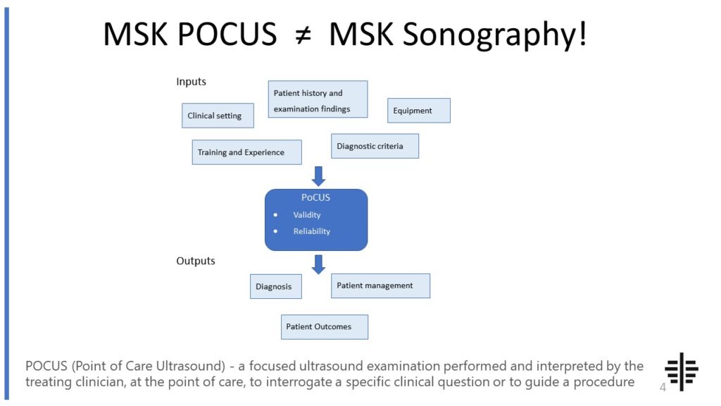
The POCUS process is very different to traditional US based in a radiology establishment. And POCUS practitioners need to be aware of those factors, unique to their particular situation, that influence diagnostic accuracy. That was the topic I presented at the plenary session of the NZAMM Annual Scientific Meeting in Wellington. A picture says 1000 ...

Posted by on 2022-10-04
We’re proud to announce that the New Zealand College of Musculoskeletal Medicine has endorsed our POCUS courses for CME and as part of vocational training. The NZCMM is responsible for setting the high standards and training of Specialist Musculoskeletal Medicine Physicians in New Zealand. NZCMM endorsement is an acknowledgement that our courses meet these standards. ...

Posted by on 2022-06-23
The RNZCUC has endorsed our courses as approved CME. We’re proud to be able to meet the training needs of Urgent Care Physicians, and look forward to meeting you at future courses.

Posted by on 2021-05-30
Ultrasound contrast agents are typically administered intravenously during a procedure. The contrast agent is injected into a vein, usually in the arm, and then circulates through the bloodstream. The timing of the injection is important, as the contrast agent needs to reach the area of interest during the ultrasound scan. The ultrasound technician will coordinate the timing of the injection with the ultrasound machine, ensuring that the contrast agent is present when the images are being captured.

Like any medical procedure, there are potential risks and side effects associated with the use of ultrasound contrast agents. The most common side effects include mild reactions at the injection site, such as pain or redness. In rare cases, more serious allergic reactions can occur, although these are extremely rare. It is important for patients to inform their healthcare provider of any known allergies or previous reactions to contrast agents. Additionally, there may be specific contraindications or precautions for certain individuals, such as pregnant women or those with kidney problems. It is important for patients to discuss their medical history with their healthcare provider before undergoing an ultrasound procedure with contrast agents.
Ultrasound contrast agents enhance the visibility of certain structures during an ultrasound scan by increasing the contrast between different tissues or blood vessels. The microbubbles or nanoparticles in the contrast agent create strong echoes when they interact with the ultrasound waves, resulting in a brighter signal. This increased signal allows for better differentiation between different structures, making it easier to identify abnormalities or specific areas of interest. By enhancing the visibility of these structures, ultrasound contrast agents can improve the accuracy and diagnostic capabilities of ultrasound imaging.

Ultrasound contrast agents can be used in a variety of ultrasound examinations, depending on the specific clinical indication. They are commonly used in echocardiography to assess the function of the heart and detect any abnormalities. Contrast agents can also be used in abdominal ultrasound examinations to improve the visualization of organs such as the liver, kidneys, or spleen. Additionally, they can be used in vascular ultrasound to assess blood flow and detect any blockages or abnormalities in the blood vessels. The use of ultrasound contrast agents is determined by the healthcare provider based on the specific clinical question and the potential benefits of using contrast.
While ultrasound contrast agents are generally safe, there are some contraindications and precautions to consider. Pregnant women are typically advised to avoid the use of contrast agents, as their safety during pregnancy has not been fully established. Individuals with severe kidney problems may also be at a higher risk of complications, as the contrast agent is eliminated through the kidneys. It is important for patients to inform their healthcare provider of any known allergies or previous reactions to contrast agents. Additionally, patients should be aware of the potential risks and benefits of using contrast agents and discuss any concerns with their healthcare provider before undergoing an ultrasound procedure.

Musculoskeletal ultrasound can be a useful tool in diagnosing myositis. This imaging technique allows for the visualization of the muscles and surrounding tissues, providing valuable information about the presence of inflammation, muscle fiber changes, and other abnormalities associated with myositis. By using high-frequency sound waves, musculoskeletal ultrasound can detect muscle edema, muscle thickening, and the presence of muscle nodules, which are characteristic features of myositis. Additionally, this imaging modality can help differentiate between different types of myositis, such as polymyositis and dermatomyositis, by assessing the pattern and distribution of muscle involvement. Overall, musculoskeletal ultrasound can play a crucial role in the diagnosis and management of myositis by providing detailed and real-time imaging of the affected muscles.
Musculoskeletal ultrasound is a valuable diagnostic tool that can accurately detect ligament tears. This imaging technique utilizes high-frequency sound waves to create detailed images of the musculoskeletal system, including ligaments. By visualizing the ligaments in real-time, musculoskeletal ultrasound can identify any abnormalities or tears present. It can provide information about the location, extent, and severity of the ligament tear, allowing healthcare professionals to make an accurate diagnosis and develop an appropriate treatment plan. Additionally, musculoskeletal ultrasound can also assess the surrounding structures and evaluate for any associated injuries or complications. Overall, musculoskeletal ultrasound is a reliable and non-invasive method for detecting ligament tears, providing valuable information for clinical decision-making.
Musculoskeletal ultrasound has been found to be an effective diagnostic tool for sacroiliitis. This imaging technique utilizes high-frequency sound waves to produce detailed images of the musculoskeletal system, including the sacroiliac joints. By visualizing the joint space, surrounding soft tissues, and any signs of inflammation or structural abnormalities, musculoskeletal ultrasound can help identify the presence of sacroiliitis. Additionally, this non-invasive and cost-effective modality allows for real-time imaging, enabling dynamic assessment of the sacroiliac joints during movement. The use of musculoskeletal ultrasound in diagnosing sacroiliitis can provide valuable information for healthcare professionals, aiding in the accurate and timely management of this condition.
Frozen shoulder syndrome, also known as adhesive capsulitis, is a condition characterized by pain and stiffness in the shoulder joint. While ultrasound is not the primary diagnostic tool for frozen shoulder syndrome, it can provide valuable information about the underlying pathology. Typical ultrasound findings in patients with frozen shoulder syndrome include thickening and inflammation of the joint capsule, as well as the presence of adhesions and fibrosis within the capsule. Additionally, ultrasound may reveal a decrease in the volume of the synovial fluid and the presence of joint effusion. These findings are indicative of the inflammatory process and the development of scar tissue within the shoulder joint, contributing to the restricted range of motion and pain experienced by patients with frozen shoulder syndrome.
Musculoskeletal ultrasound plays a crucial role in diagnosing carpal tunnel syndrome by providing detailed imaging of the musculoskeletal structures in the wrist and hand. This non-invasive imaging technique allows healthcare professionals to visualize the median nerve, tendons, ligaments, and surrounding tissues in real-time. By assessing the size and shape of the median nerve, as well as any abnormalities such as swelling or compression, musculoskeletal ultrasound can help confirm the presence of carpal tunnel syndrome. Additionally, this imaging modality can also identify other potential causes of symptoms, such as tendonitis or ganglion cysts, ensuring an accurate diagnosis and appropriate treatment plan.