
Ultrasound plays a crucial role in diagnosing referred pain syndromes by providing real-time imaging of the affected area. It allows healthcare professionals to visualize the soft tissues, muscles, tendons, and nerves, which can help identify any abnormalities or sources of pain. By using ultrasound, doctors can assess the structure and function of the affected area, determine the extent of any damage or inflammation, and pinpoint the exact location of the referred pain. This imaging technique helps in differentiating between referred pain and other potential causes, enabling accurate diagnosis and appropriate treatment planning.
There are several common referred pain syndromes that can be detected using ultrasound. One such syndrome is myofascial pain syndrome, which is characterized by trigger points in the muscles that refer pain to other areas of the body. Ultrasound can identify these trigger points and assess the surrounding tissues for any abnormalities. Another common syndrome is radiculopathy, where nerve compression or irritation leads to pain radiating along the nerve pathway. Ultrasound can visualize the affected nerve roots and assess for any compression or inflammation. Additionally, ultrasound can help diagnose referred pain syndromes related to joint dysfunction, such as hip or shoulder impingement, by visualizing the joint structures and assessing for any abnormalities.
Over the last couple of years, we’ve brought you several courses focusing on Ultrasound Guided Injection Techniques. They’ve been extremely popular, and like our other courses, the feedback has been fantastic. One thing we’ve learnt along the way is that to get the most out of learning injection techniques, a solid grounding in MSK Ultrasound ...
Posted by on 2024-02-10
What a year 2023 was! We’ve loved bringing you courses covering US of the upper and lower limb, and US guided injections through the year. The mix of health professionals from all sorts of backgrounds (Doctors, Nurses, Physios, Sonographers to name a few) has been amazing to be part of. We’ve been humbled by your ...
Posted by on 2023-09-17
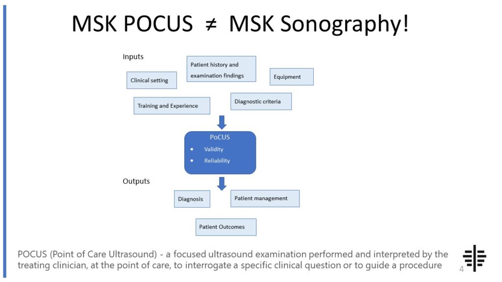
The POCUS process is very different to traditional US based in a radiology establishment. And POCUS practitioners need to be aware of those factors, unique to their particular situation, that influence diagnostic accuracy. That was the topic I presented at the plenary session of the NZAMM Annual Scientific Meeting in Wellington. A picture says 1000 ...

Posted by on 2022-10-04
We’re proud to announce that the New Zealand College of Musculoskeletal Medicine has endorsed our POCUS courses for CME and as part of vocational training. The NZCMM is responsible for setting the high standards and training of Specialist Musculoskeletal Medicine Physicians in New Zealand. NZCMM endorsement is an acknowledgement that our courses meet these standards. ...

Posted by on 2022-06-23
The RNZCUC has endorsed our courses as approved CME. We’re proud to be able to meet the training needs of Urgent Care Physicians, and look forward to meeting you at future courses.

Posted by on 2021-05-30
Ultrasound can provide valuable information to differentiate between referred pain syndromes and other musculoskeletal conditions. However, it is important to note that ultrasound findings should be interpreted in conjunction with the patient's clinical history and physical examination. While ultrasound can visualize soft tissues and identify potential sources of referred pain, it may not always provide a definitive diagnosis. In some cases, further imaging modalities or diagnostic tests may be required to confirm the diagnosis or rule out other conditions. Therefore, ultrasound should be used as a complementary tool in the diagnostic process, along with other clinical assessments.

There are several advantages of using ultrasound over other imaging modalities for evaluating referred pain syndromes. Firstly, ultrasound is a non-invasive and radiation-free imaging technique, making it safe for repeated examinations and suitable for patients of all ages. It provides real-time imaging, allowing for dynamic assessment of the affected area and the ability to reproduce the patient's pain during the examination. Ultrasound is also cost-effective compared to other imaging modalities such as MRI or CT scans. Additionally, ultrasound can be performed at the point of care, allowing for immediate diagnosis and treatment planning, which can be particularly beneficial in acute or urgent cases.
Ultrasound-guided intervention plays a crucial role in managing referred pain syndromes. By using ultrasound to visualize the affected area in real-time, healthcare professionals can accurately guide injections or other therapeutic procedures to the precise location of the pain source. This ensures that the treatment is targeted and maximizes its effectiveness. Ultrasound guidance also reduces the risk of complications by avoiding vital structures and improving the accuracy of needle placement. It allows for real-time monitoring of the procedure, ensuring that the medication or intervention is delivered to the intended site, providing immediate relief and improving patient outcomes.

While ultrasound is generally safe and well-tolerated, there are some limitations and contraindications to its use for referred pain syndromes. Ultrasound may not be suitable for patients with significant obesity or excessive tissue thickness, as it may limit the penetration of sound waves and affect image quality. Additionally, certain anatomical locations may be challenging to visualize using ultrasound, such as deep-seated structures or areas with limited acoustic windows. In some cases, patients with metal implants or devices may not be suitable candidates for ultrasound due to potential interference or artifacts. It is important for healthcare professionals to assess each patient's individual circumstances and determine the appropriateness of ultrasound for their specific case.
The potential complications or risks associated with ultrasound-guided interventions for referred pain syndromes are generally minimal. However, as with any medical procedure, there is a small risk of infection, bleeding, or allergic reactions to medications used during the intervention. These risks can be minimized by following proper sterile techniques and using appropriate precautions. Additionally, there may be a slight discomfort or pain during the procedure, but this is usually temporary and well-tolerated. It is important for healthcare professionals to discuss the potential risks and benefits of ultrasound-guided interventions with the patient and obtain informed consent before proceeding with the procedure.

Ultrasound offers several advantages over MRI for diagnosing musculoskeletal disorders. Firstly, ultrasound is a non-invasive and painless imaging technique that does not involve exposure to ionizing radiation, making it a safer option for patients, especially those who may require multiple imaging sessions. Additionally, ultrasound provides real-time imaging, allowing for dynamic assessment of the musculoskeletal system during movement or stress tests. This real-time capability enables the visualization of soft tissues, such as tendons, ligaments, and muscles, in their natural state, providing valuable information about their structure and function. Moreover, ultrasound is more cost-effective and readily available compared to MRI, making it a more accessible diagnostic tool for musculoskeletal disorders. Overall, the use of ultrasound in diagnosing musculoskeletal disorders offers numerous benefits, including safety, real-time imaging, and cost-effectiveness.
Musculoskeletal ultrasound plays a crucial role in the evaluation of synovial sarcoma by providing valuable information about the tumor's location, size, and characteristics. This imaging technique utilizes high-frequency sound waves to create detailed images of the soft tissues and structures surrounding the affected area. By examining the synovial sarcoma with ultrasound, healthcare professionals can assess the tumor's extent of infiltration into adjacent tissues, identify any associated cystic or necrotic areas, and determine the presence of vascular involvement. Additionally, musculoskeletal ultrasound allows for real-time visualization, enabling the evaluation of dynamic changes in the tumor during movement or manipulation. This non-invasive and cost-effective imaging modality aids in the accurate diagnosis, staging, and monitoring of synovial sarcoma, ultimately guiding treatment decisions and improving patient outcomes.