
Ultrasound-guided injection improves the accuracy of needle placement by providing real-time visualization of the target area. The ultrasound machine uses sound waves to create images of the internal structures, allowing the healthcare professional to see the exact location of the needle in relation to the target. This eliminates the need for blind injections and reduces the risk of complications such as accidental puncture of blood vessels or nerves. The ability to visualize the needle in real-time also allows for adjustments to be made during the injection, ensuring precise placement and increasing the likelihood of successful outcomes.
There are several advantages of using ultrasound guidance for injections compared to other techniques. Firstly, it provides real-time visualization, allowing for accurate needle placement and reducing the risk of complications. Secondly, it can be used to guide injections in difficult-to-reach areas or areas with complex anatomy, such as joints, tendons, or nerves. Thirdly, ultrasound guidance allows for the visualization of structures that may not be visible with other techniques, such as small blood vessels or nerves. This can help avoid accidental damage during the injection. Overall, ultrasound-guided injections offer improved accuracy, increased safety, and the ability to target specific structures with precision.
Over the last couple of years, we’ve brought you several courses focusing on Ultrasound Guided Injection Techniques. They’ve been extremely popular, and like our other courses, the feedback has been fantastic. One thing we’ve learnt along the way is that to get the most out of learning injection techniques, a solid grounding in MSK Ultrasound ...
Posted by on 2024-02-10
What a year 2023 was! We’ve loved bringing you courses covering US of the upper and lower limb, and US guided injections through the year. The mix of health professionals from all sorts of backgrounds (Doctors, Nurses, Physios, Sonographers to name a few) has been amazing to be part of. We’ve been humbled by your ...
Posted by on 2023-09-17
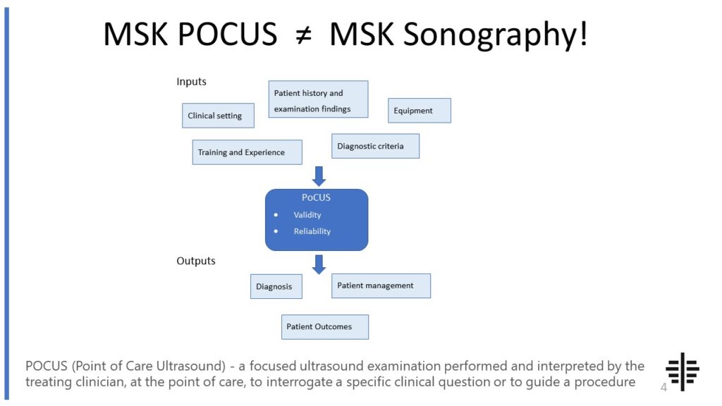
The POCUS process is very different to traditional US based in a radiology establishment. And POCUS practitioners need to be aware of those factors, unique to their particular situation, that influence diagnostic accuracy. That was the topic I presented at the plenary session of the NZAMM Annual Scientific Meeting in Wellington. A picture says 1000 ...

Posted by on 2022-10-04
We’re proud to announce that the New Zealand College of Musculoskeletal Medicine has endorsed our POCUS courses for CME and as part of vocational training. The NZCMM is responsible for setting the high standards and training of Specialist Musculoskeletal Medicine Physicians in New Zealand. NZCMM endorsement is an acknowledgement that our courses meet these standards. ...

Posted by on 2022-06-23
Ultrasound-guided injections can be used for a wide range of injections, including joint injections, nerve blocks, tendon injections, and soft tissue injections. The technique is particularly effective for injections in areas with complex anatomy or structures that are difficult to visualize, such as deep joints or small nerves. It can also be beneficial for injections that require precise needle placement, such as epidural injections or injections into specific muscle groups. However, the use of ultrasound guidance may not be necessary for all types of injections, especially those that can be easily performed using other techniques with high accuracy.

Like any medical procedure, there are potential risks and complications associated with ultrasound-guided injections. These can include infection at the injection site, bleeding, nerve damage, or allergic reactions to the injected medication. However, the use of ultrasound guidance can actually help reduce these risks by providing real-time visualization and precise needle placement. Additionally, healthcare professionals who perform ultrasound-guided injections should have proper training and experience to minimize the risk of complications. It is important for patients to discuss any potential risks or concerns with their healthcare provider before undergoing an ultrasound-guided injection.
The use of ultrasound guidance significantly improves the success rate of injections. By providing real-time visualization and precise needle placement, ultrasound-guided injections increase the accuracy of delivering the medication to the intended target. This can result in improved pain relief, better outcomes, and reduced need for repeat injections. Studies have shown that ultrasound-guided injections have higher success rates compared to blind injections or injections guided by other techniques. The ability to visualize the needle in real-time allows for adjustments to be made during the injection, ensuring that the medication is delivered to the desired location.

While ultrasound-guided injections offer many benefits, there are some limitations to consider. Firstly, the technique requires specialized equipment and training, which may not be readily available in all healthcare settings. Additionally, ultrasound-guided injections may take longer to perform compared to blind injections or injections guided by other techniques. This can be a limitation in certain situations where time is a critical factor. Lastly, the effectiveness of ultrasound-guided injections may depend on the skill and experience of the healthcare professional performing the procedure. Proper training and proficiency in ultrasound imaging are essential to ensure accurate needle placement and maximize the benefits of this technique.
Healthcare professionals who perform ultrasound-guided injections should undergo specific training and certification to ensure competency in the technique. This typically involves completing a specialized course or program that covers the principles of ultrasound imaging, anatomy, needle guidance, and injection techniques. Certification may be obtained through professional organizations or societies that offer ultrasound-guided injection training programs. It is important for healthcare professionals to stay updated with the latest advancements and guidelines in ultrasound-guided injections to provide safe and effective care to their patients.

Musculoskeletal ultrasound plays a crucial role in the diagnosis of adhesive capsulitis by providing detailed imaging of the affected joint and surrounding structures. This imaging technique allows for the visualization of the thickening and inflammation of the joint capsule, which are characteristic features of adhesive capsulitis. Additionally, musculoskeletal ultrasound can help identify any associated abnormalities such as bursitis or tendonitis that may be contributing to the symptoms. By accurately assessing the extent and location of the pathology, musculoskeletal ultrasound aids in differentiating adhesive capsulitis from other conditions with similar clinical presentations. Furthermore, this diagnostic tool enables real-time assessment of joint mobility and can be used to guide therapeutic interventions such as corticosteroid injections or physical therapy. Overall, musculoskeletal ultrasound is a valuable tool in the diagnosis and management of adhesive capsulitis, providing clinicians with essential information to develop an appropriate treatment plan.
Musculoskeletal ultrasound has a wide range of applications in sports medicine. It is commonly used for the diagnosis and monitoring of various musculoskeletal injuries and conditions. For example, it can be used to assess the extent and severity of muscle strains, tendonitis, and ligament injuries. It can also be used to evaluate joint inflammation, such as in cases of arthritis or bursitis. Additionally, musculoskeletal ultrasound can be used to guide therapeutic injections, such as corticosteroid injections, into specific areas of the body for pain relief and inflammation reduction. This imaging technique is also valuable for assessing the healing progress of injuries and monitoring the effectiveness of rehabilitation programs. Overall, musculoskeletal ultrasound plays a crucial role in the accurate diagnosis, treatment, and management of musculoskeletal conditions in athletes and individuals involved in sports activities.
Calcific tendinitis is a condition characterized by the deposition of calcium crystals within the tendons, most commonly affecting the rotator cuff tendons in the shoulder. Sonographic imaging plays a crucial role in the diagnosis of calcific tendinitis, as it allows for the visualization of the characteristic features associated with this condition. On ultrasound, calcific tendinitis appears as hyperechoic foci within the affected tendon, representing the calcific deposits. These foci may exhibit variable echogenicity, ranging from punctate to linear or even curvilinear patterns. The size and shape of the calcific deposits can also vary, with some appearing as small, discrete foci and others forming larger, irregular masses. Additionally, the presence of acoustic shadowing posterior to the calcific deposits is a common finding, further aiding in the diagnosis. Overall, sonographic features of calcific tendinitis include hyperechoic foci with variable echogenicity, variable size and shape, and the presence of acoustic shadowing.
Musculoskeletal ultrasound is a valuable imaging modality that can aid in the diagnosis of stress fractures. This non-invasive technique utilizes high-frequency sound waves to produce detailed images of the musculoskeletal system, allowing for the visualization of bone structures and surrounding soft tissues. By assessing the bone cortex, periosteum, and adjacent soft tissues, musculoskeletal ultrasound can help identify the characteristic signs of stress fractures, such as cortical irregularities, periosteal reactions, and localized edema. Additionally, this imaging technique can provide real-time dynamic assessment, allowing for the detection of stress fracture-related changes during movement or weight-bearing activities. While musculoskeletal ultrasound is a useful tool in the diagnosis of stress fractures, it is often used in conjunction with other imaging modalities, such as X-ray or magnetic resonance imaging (MRI), to ensure accurate and comprehensive evaluation.
Musculoskeletal ultrasound plays a crucial role in the diagnosis of Morton's neuroma by providing detailed imaging of the affected area. This non-invasive imaging technique utilizes high-frequency sound waves to create real-time images of the musculoskeletal structures, including the foot. By using musculoskeletal ultrasound, healthcare professionals can visualize the neuroma, a benign growth of nerve tissue, in the intermetatarsal spaces of the foot. The ultrasound can accurately identify the size, location, and extent of the neuroma, allowing for a precise diagnosis. Additionally, musculoskeletal ultrasound can help differentiate Morton's neuroma from other conditions that may present with similar symptoms, such as stress fractures or bursitis. Overall, musculoskeletal ultrasound aids in the diagnosis of Morton's neuroma by providing valuable visual information that assists healthcare professionals in making an accurate and timely diagnosis.
Musculoskeletal ultrasound has the potential to differentiate between benign and malignant soft tissue tumors. This imaging technique utilizes high-frequency sound waves to produce detailed images of the musculoskeletal system, allowing for the evaluation of various soft tissue abnormalities. By assessing the characteristics of the tumor, such as size, shape, vascularity, and internal architecture, musculoskeletal ultrasound can provide valuable information that aids in distinguishing between benign and malignant tumors. Additionally, the use of Doppler ultrasound can assess blood flow within the tumor, which can be indicative of malignancy. However, it is important to note that while musculoskeletal ultrasound can provide valuable information, it is not always definitive in differentiating between benign and malignant soft tissue tumors. Therefore, further diagnostic tests, such as biopsy or magnetic resonance imaging (MRI), may be necessary for a conclusive diagnosis.