
Ultrasound can be used to monitor disease progression by providing real-time imaging of internal organs and tissues. It uses high-frequency sound waves to create detailed images of the body, allowing healthcare professionals to visualize any changes or abnormalities. For example, in the case of cancer, ultrasound can be used to monitor the size and growth of tumors over time. By regularly performing ultrasound scans, doctors can track the progression of the disease and make informed decisions about treatment options.
There are several advantages of using ultrasound for disease monitoring compared to other imaging techniques. Firstly, ultrasound is non-invasive and does not involve the use of ionizing radiation, making it a safer option for patients. Additionally, ultrasound is relatively inexpensive and widely available, making it more accessible for routine monitoring. Furthermore, ultrasound provides real-time imaging, allowing healthcare professionals to immediately visualize any changes or developments in the disease progression. This real-time information can be crucial in making timely treatment decisions.
Over the last couple of years, we’ve brought you several courses focusing on Ultrasound Guided Injection Techniques. They’ve been extremely popular, and like our other courses, the feedback has been fantastic. One thing we’ve learnt along the way is that to get the most out of learning injection techniques, a solid grounding in MSK Ultrasound ...
Posted by on 2024-02-10
What a year 2023 was! We’ve loved bringing you courses covering US of the upper and lower limb, and US guided injections through the year. The mix of health professionals from all sorts of backgrounds (Doctors, Nurses, Physios, Sonographers to name a few) has been amazing to be part of. We’ve been humbled by your ...
Posted by on 2023-09-17
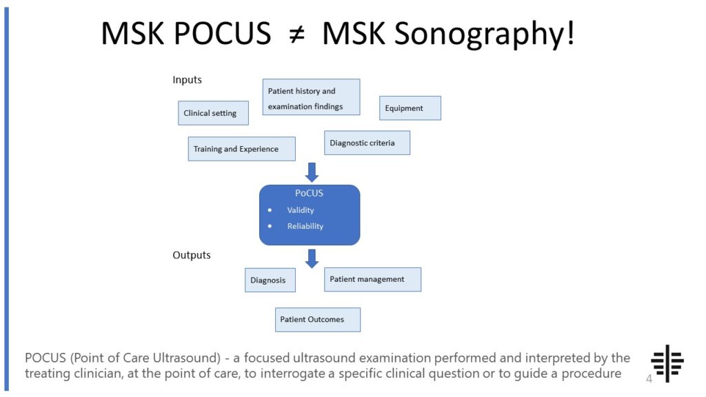
The POCUS process is very different to traditional US based in a radiology establishment. And POCUS practitioners need to be aware of those factors, unique to their particular situation, that influence diagnostic accuracy. That was the topic I presented at the plenary session of the NZAMM Annual Scientific Meeting in Wellington. A picture says 1000 ...

Posted by on 2022-10-04
We’re proud to announce that the New Zealand College of Musculoskeletal Medicine has endorsed our POCUS courses for CME and as part of vocational training. The NZCMM is responsible for setting the high standards and training of Specialist Musculoskeletal Medicine Physicians in New Zealand. NZCMM endorsement is an acknowledgement that our courses meet these standards. ...

Posted by on 2022-06-23
The RNZCUC has endorsed our courses as approved CME. We’re proud to be able to meet the training needs of Urgent Care Physicians, and look forward to meeting you at future courses.

Posted by on 2021-05-30
Ultrasound can effectively monitor a wide range of diseases across various medical specialties. In cardiology, it can be used to monitor heart conditions such as heart failure, valve abnormalities, and congenital heart defects. In obstetrics and gynecology, ultrasound is commonly used to monitor the development of the fetus during pregnancy and detect any abnormalities. It is also used in the diagnosis and monitoring of liver diseases, kidney diseases, and musculoskeletal conditions. Overall, ultrasound is a versatile imaging technique that can be applied to monitor diseases in multiple organ systems.

Ultrasound provides real-time information about disease progression by using sound waves to create live images of the body. The ultrasound machine sends high-frequency sound waves into the body, which then bounce back and create echoes. These echoes are captured by the machine and converted into images that can be viewed in real-time. This allows healthcare professionals to observe any changes or developments in the disease progression immediately. For example, during an ultrasound scan of a tumor, doctors can see if the tumor has grown in size or if there are any new abnormalities present.
Yes, ultrasound can be used to detect early signs of disease progression. Due to its ability to provide real-time imaging, ultrasound can capture subtle changes in the body that may indicate the early stages of a disease. For example, in the case of liver diseases, ultrasound can detect the presence of fatty liver or early signs of liver fibrosis. Similarly, in the case of cardiovascular diseases, ultrasound can detect the thickening of arterial walls or the presence of plaque, which are early indicators of atherosclerosis. Early detection through ultrasound can lead to timely interventions and improved outcomes.

While ultrasound is a valuable tool for disease monitoring, it does have some limitations and drawbacks. One limitation is that ultrasound is highly operator-dependent, meaning that the quality of the images can vary depending on the skill and experience of the person performing the scan. Additionally, ultrasound may not be suitable for imaging certain areas of the body, such as the lungs or bones, as sound waves do not penetrate these structures well. Furthermore, ultrasound may not provide as detailed images as other imaging techniques like MRI or CT scans, which can limit its ability to detect certain diseases or abnormalities.
Ultrasound has been used in various clinical settings to monitor disease progression. For example, in oncology, ultrasound is used to monitor the growth and spread of tumors, assess the response to treatment, and guide biopsies. In cardiology, ultrasound is used to monitor heart function, assess the presence of heart defects, and guide procedures such as cardiac catheterization. In obstetrics, ultrasound is used to monitor fetal development, detect any abnormalities, and guide interventions if necessary. These are just a few examples of how ultrasound has been successfully utilized in clinical practice to monitor disease progression and guide patient management.

Musculoskeletal ultrasound has a wide range of applications in sports medicine. It is commonly used for the diagnosis and monitoring of various musculoskeletal injuries and conditions. For example, it can be used to assess the extent and severity of muscle strains, tendonitis, and ligament injuries. It can also be used to evaluate joint inflammation, such as in cases of arthritis or bursitis. Additionally, musculoskeletal ultrasound can be used to guide therapeutic injections, such as corticosteroid injections, into specific areas of the body for pain relief and inflammation reduction. This imaging technique is also valuable for assessing the healing progress of injuries and monitoring the effectiveness of rehabilitation programs. Overall, musculoskeletal ultrasound plays a crucial role in the accurate diagnosis, treatment, and management of musculoskeletal conditions in athletes and individuals involved in sports activities.
Calcific tendinitis is a condition characterized by the deposition of calcium crystals within the tendons, most commonly affecting the rotator cuff tendons in the shoulder. Sonographic imaging plays a crucial role in the diagnosis of calcific tendinitis, as it allows for the visualization of the characteristic features associated with this condition. On ultrasound, calcific tendinitis appears as hyperechoic foci within the affected tendon, representing the calcific deposits. These foci may exhibit variable echogenicity, ranging from punctate to linear or even curvilinear patterns. The size and shape of the calcific deposits can also vary, with some appearing as small, discrete foci and others forming larger, irregular masses. Additionally, the presence of acoustic shadowing posterior to the calcific deposits is a common finding, further aiding in the diagnosis. Overall, sonographic features of calcific tendinitis include hyperechoic foci with variable echogenicity, variable size and shape, and the presence of acoustic shadowing.
Musculoskeletal ultrasound is a valuable imaging modality that can aid in the diagnosis of stress fractures. This non-invasive technique utilizes high-frequency sound waves to produce detailed images of the musculoskeletal system, allowing for the visualization of bone structures and surrounding soft tissues. By assessing the bone cortex, periosteum, and adjacent soft tissues, musculoskeletal ultrasound can help identify the characteristic signs of stress fractures, such as cortical irregularities, periosteal reactions, and localized edema. Additionally, this imaging technique can provide real-time dynamic assessment, allowing for the detection of stress fracture-related changes during movement or weight-bearing activities. While musculoskeletal ultrasound is a useful tool in the diagnosis of stress fractures, it is often used in conjunction with other imaging modalities, such as X-ray or magnetic resonance imaging (MRI), to ensure accurate and comprehensive evaluation.
Musculoskeletal ultrasound plays a crucial role in the diagnosis of Morton's neuroma by providing detailed imaging of the affected area. This non-invasive imaging technique utilizes high-frequency sound waves to create real-time images of the musculoskeletal structures, including the foot. By using musculoskeletal ultrasound, healthcare professionals can visualize the neuroma, a benign growth of nerve tissue, in the intermetatarsal spaces of the foot. The ultrasound can accurately identify the size, location, and extent of the neuroma, allowing for a precise diagnosis. Additionally, musculoskeletal ultrasound can help differentiate Morton's neuroma from other conditions that may present with similar symptoms, such as stress fractures or bursitis. Overall, musculoskeletal ultrasound aids in the diagnosis of Morton's neuroma by providing valuable visual information that assists healthcare professionals in making an accurate and timely diagnosis.
Musculoskeletal ultrasound has the potential to differentiate between benign and malignant soft tissue tumors. This imaging technique utilizes high-frequency sound waves to produce detailed images of the musculoskeletal system, allowing for the evaluation of various soft tissue abnormalities. By assessing the characteristics of the tumor, such as size, shape, vascularity, and internal architecture, musculoskeletal ultrasound can provide valuable information that aids in distinguishing between benign and malignant tumors. Additionally, the use of Doppler ultrasound can assess blood flow within the tumor, which can be indicative of malignancy. However, it is important to note that while musculoskeletal ultrasound can provide valuable information, it is not always definitive in differentiating between benign and malignant soft tissue tumors. Therefore, further diagnostic tests, such as biopsy or magnetic resonance imaging (MRI), may be necessary for a conclusive diagnosis.