
Ultrasound therapy is beneficial in the treatment of recurrent injuries as it helps to promote tissue healing and reduce inflammation. The high-frequency sound waves generated by the ultrasound machine penetrate deep into the injured tissues, stimulating blood flow and increasing the delivery of oxygen and nutrients to the area. This enhanced circulation helps to accelerate the healing process and reduce pain. Additionally, ultrasound therapy can also break down scar tissue and adhesions, which can contribute to recurrent injuries by limiting range of motion and causing further damage.
There are several mechanisms through which ultrasound promotes healing in recurrent injuries. Firstly, the mechanical vibrations produced by the sound waves can stimulate the production of collagen, a protein that is essential for tissue repair. This can help to strengthen the injured area and prevent future injuries. Secondly, ultrasound therapy can increase the permeability of cell membranes, allowing for better absorption of medications or other therapeutic substances that may be applied topically. This can further enhance the healing process and reduce the risk of recurrent injuries.
Over the last couple of years, we’ve brought you several courses focusing on Ultrasound Guided Injection Techniques. They’ve been extremely popular, and like our other courses, the feedback has been fantastic. One thing we’ve learnt along the way is that to get the most out of learning injection techniques, a solid grounding in MSK Ultrasound ...
Posted by on 2024-02-10
What a year 2023 was! We’ve loved bringing you courses covering US of the upper and lower limb, and US guided injections through the year. The mix of health professionals from all sorts of backgrounds (Doctors, Nurses, Physios, Sonographers to name a few) has been amazing to be part of. We’ve been humbled by your ...
Posted by on 2023-09-17
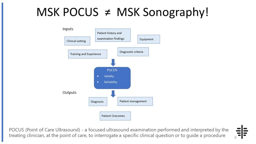
The POCUS process is very different to traditional US based in a radiology establishment. And POCUS practitioners need to be aware of those factors, unique to their particular situation, that influence diagnostic accuracy. That was the topic I presented at the plenary session of the NZAMM Annual Scientific Meeting in Wellington. A picture says 1000 ...

Posted by on 2022-10-04
We’re proud to announce that the New Zealand College of Musculoskeletal Medicine has endorsed our POCUS courses for CME and as part of vocational training. The NZCMM is responsible for setting the high standards and training of Specialist Musculoskeletal Medicine Physicians in New Zealand. NZCMM endorsement is an acknowledgement that our courses meet these standards. ...

Posted by on 2022-06-23
The RNZCUC has endorsed our courses as approved CME. We’re proud to be able to meet the training needs of Urgent Care Physicians, and look forward to meeting you at future courses.

Posted by on 2021-05-30
While ultrasound therapy is primarily used as a treatment for recurrent injuries, it can also be used as a preventive measure to reduce the risk of future injuries. By promoting tissue healing and strengthening the injured area, ultrasound therapy can help to improve the overall stability and resilience of the tissues. This can make them less susceptible to reinjury and reduce the likelihood of recurrent injuries. However, it is important to note that ultrasound therapy should be used in conjunction with other preventive measures, such as proper warm-up exercises and appropriate training techniques, for optimal results.

Ultrasound therapy has been found to be effective in treating a wide range of recurrent injuries, including tendinitis, muscle strains, ligament sprains, and bursitis. However, the specific types of injuries that respond better to ultrasound therapy may vary depending on various factors, such as the severity and location of the injury, as well as the individual's overall health and healing capacity. It is recommended to consult with a healthcare professional to determine if ultrasound therapy is appropriate for a specific recurrent injury.
The effectiveness of ultrasound therapy compared to other treatment options for recurrent injuries can vary depending on the individual and the specific injury. While ultrasound therapy has been shown to be effective in promoting tissue healing and reducing pain, it may not be the most suitable option for all cases. Other treatment options, such as physical therapy, medication, or surgery, may be more appropriate depending on the nature and severity of the injury. It is important to consult with a healthcare professional to determine the most effective treatment approach for a specific recurrent injury.

Generally, ultrasound therapy is considered to be a safe and non-invasive treatment option for recurrent injuries. However, there are some potential side effects and risks associated with its use. These can include mild discomfort or pain during the treatment, skin irritation or burns if the ultrasound probe is not properly applied, and the potential for exacerbating an underlying condition if the therapy is not administered correctly. It is important to follow the guidance of a healthcare professional and ensure that the ultrasound therapy is performed by a trained and qualified practitioner to minimize the risk of side effects or complications.
The frequency and duration of ultrasound therapy sessions for treating recurrent injuries can vary depending on the specific injury and the individual's response to treatment. Typically, ultrasound therapy sessions are scheduled 2-3 times per week, with each session lasting around 5-10 minutes. However, the exact frequency and duration may be adjusted based on the individual's progress and the healthcare professional's assessment. It is important to follow the recommended treatment plan and attend all scheduled sessions to maximize the benefits of ultrasound therapy for recurrent injuries.

Musculoskeletal ultrasound has emerged as a valuable tool for assessing elbow pathology; however, it is not without its challenges. One of the main challenges is the limited field of view provided by ultrasound imaging, which can make it difficult to visualize the entire elbow joint and surrounding structures. Additionally, the complex anatomy of the elbow, including the presence of multiple tendons, ligaments, and bony structures, can pose challenges in accurately identifying and differentiating between various pathologies. Furthermore, the operator's skill and experience in performing musculoskeletal ultrasound plays a crucial role in obtaining high-quality images and interpreting them correctly. The dynamic nature of the elbow joint, with its wide range of motion, can also make it challenging to capture images in real-time and accurately assess the pathology. Despite these challenges, advancements in technology and ongoing research in musculoskeletal ultrasound continue to improve its utility in assessing elbow pathology.
Musculoskeletal ultrasound is a valuable imaging modality that can aid in the identification of Baker's cysts. Baker's cysts, also known as popliteal cysts, are fluid-filled sacs that develop in the posterior aspect of the knee joint. These cysts are often associated with underlying knee joint pathology, such as osteoarthritis or meniscal tears. When performing a musculoskeletal ultrasound, the sonographer can utilize high-frequency sound waves to visualize the cyst and assess its characteristics. This imaging technique allows for the identification of the cyst's location, size, shape, and internal content. Additionally, musculoskeletal ultrasound can help differentiate Baker's cysts from other knee joint abnormalities, such as synovial cysts or tumors. By employing musculoskeletal ultrasound, healthcare professionals can accurately diagnose and monitor the presence of Baker's cysts, facilitating appropriate management and treatment decisions.
Musculoskeletal ultrasound plays a crucial role in the evaluation of complex regional pain syndrome (CRPS) by providing valuable insights into the underlying pathophysiology and aiding in the diagnosis and management of this condition. By utilizing high-frequency sound waves, musculoskeletal ultrasound allows for the visualization of soft tissues, joints, and nerves, enabling the identification of structural abnormalities, such as edema, synovitis, and nerve entrapment. This imaging modality also facilitates the assessment of blood flow dynamics, which is particularly relevant in CRPS, as vascular dysfunction is a key feature of the condition. Additionally, musculoskeletal ultrasound can guide interventions, such as nerve blocks and injections, providing targeted and precise treatment options for patients with CRPS. Overall, musculoskeletal ultrasound serves as a valuable tool in the comprehensive evaluation and management of complex regional pain syndrome.
Typical findings in musculoskeletal ultrasound of patients with osteoporosis may include decreased bone density, cortical thinning, and increased echogenicity of the trabecular bone. The ultrasound may also reveal the presence of osteophytes, joint effusion, and synovial thickening. Additionally, the ultrasound may show signs of muscle atrophy and fatty infiltration, as well as tendon abnormalities such as tendon thickening or tears. These findings are indicative of the structural changes that occur in the musculoskeletal system due to osteoporosis, highlighting the importance of ultrasound in the assessment and management of this condition.
Musculoskeletal ultrasound is a valuable tool for assessing sacroiliac joint dysfunction, but it does have some limitations. One limitation is that it can be challenging to obtain clear and accurate images of the sacroiliac joint due to its deep location and the presence of overlying structures such as muscles and ligaments. Additionally, the interpretation of ultrasound images can be subjective and dependent on the experience and expertise of the operator. Another limitation is that ultrasound may not be able to provide a comprehensive assessment of the sacroiliac joint, as it may not be able to visualize certain structures such as the articular cartilage or the joint space. Furthermore, ultrasound is limited in its ability to assess the functional aspects of the sacroiliac joint, such as joint mobility or stability. Therefore, while musculoskeletal ultrasound can be a useful tool in the assessment of sacroiliac joint dysfunction, it should be used in conjunction with other imaging modalities and clinical findings to ensure a comprehensive evaluation.
Musculoskeletal ultrasound can be a useful tool for diagnosing vertebral compression fractures. This imaging technique utilizes high-frequency sound waves to create detailed images of the musculoskeletal system, including the spine. By examining the affected area, musculoskeletal ultrasound can help identify signs of vertebral compression fractures, such as changes in bone density, deformities, or the presence of fractures. Additionally, musculoskeletal ultrasound can provide real-time visualization, allowing for dynamic assessment of the spine during movement or weight-bearing activities. This can aid in the accurate diagnosis and monitoring of vertebral compression fractures, guiding appropriate treatment strategies.