
Ultrasonographic measurement techniques are based on the principle of using high-frequency sound waves to create images of internal structures in the body. These sound waves are emitted by a transducer and then bounce back when they encounter different tissues or structures. The transducer detects the returning sound waves and converts them into electrical signals, which are then processed to create a visual representation of the scanned area. By analyzing the echoes produced by the sound waves, ultrasonographic measurement techniques can provide information about the size, shape, and composition of organs, tissues, and other structures within the body.
Ultrasonographic measurement differs from other imaging modalities in several ways. Firstly, it is non-invasive, meaning it does not require any incisions or injections. This makes it a safer and more comfortable option for patients. Additionally, ultrasonography does not use ionizing radiation, unlike X-rays or CT scans, making it a preferred choice for pregnant women and children. Furthermore, ultrasonographic measurement provides real-time imaging, allowing for dynamic assessment of structures and functions. It is also portable and relatively inexpensive compared to other imaging techniques, making it more accessible in various healthcare settings.
Over the last couple of years, we’ve brought you several courses focusing on Ultrasound Guided Injection Techniques. They’ve been extremely popular, and like our other courses, the feedback has been fantastic. One thing we’ve learnt along the way is that to get the most out of learning injection techniques, a solid grounding in MSK Ultrasound ...
Posted by on 2024-02-10
What a year 2023 was! We’ve loved bringing you courses covering US of the upper and lower limb, and US guided injections through the year. The mix of health professionals from all sorts of backgrounds (Doctors, Nurses, Physios, Sonographers to name a few) has been amazing to be part of. We’ve been humbled by your ...
Posted by on 2023-09-17
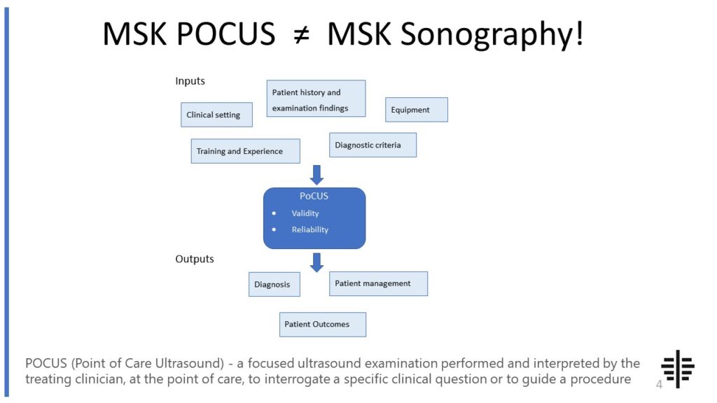
The POCUS process is very different to traditional US based in a radiology establishment. And POCUS practitioners need to be aware of those factors, unique to their particular situation, that influence diagnostic accuracy. That was the topic I presented at the plenary session of the NZAMM Annual Scientific Meeting in Wellington. A picture says 1000 ...

Posted by on 2022-10-04
We’re proud to announce that the New Zealand College of Musculoskeletal Medicine has endorsed our POCUS courses for CME and as part of vocational training. The NZCMM is responsible for setting the high standards and training of Specialist Musculoskeletal Medicine Physicians in New Zealand. NZCMM endorsement is an acknowledgement that our courses meet these standards. ...

Posted by on 2022-06-23
There are several advantages to using ultrasonographic measurement techniques. Firstly, it is a safe and non-invasive procedure that does not expose patients to ionizing radiation. This makes it suitable for repeated examinations and for vulnerable populations such as pregnant women and children. Secondly, ultrasonography provides real-time imaging, allowing for immediate visualization and assessment of structures and functions. It is also a versatile technique that can be used to examine various organs and systems in the body. Additionally, ultrasonographic measurement is relatively inexpensive and widely available, making it a cost-effective option for medical imaging.

Ultrasonographic measurement techniques have numerous applications in medical imaging. One common application is in obstetrics, where it is used to monitor fetal growth and development, assess the placenta, and detect any abnormalities or complications during pregnancy. It is also used in cardiology to evaluate the structure and function of the heart, including the valves and blood flow. In gastroenterology, ultrasonography can be used to examine the liver, gallbladder, pancreas, and other abdominal organs. It is also used in urology to assess the kidneys, bladder, and prostate. Additionally, ultrasonography is used in musculoskeletal imaging to evaluate joints, tendons, and muscles.
Ultrasonographic measurement is commonly used in assessing fetal growth and development. It allows healthcare providers to measure various parameters such as the size of the fetus, the position of the placenta, and the amount of amniotic fluid. It can also be used to assess the fetal heart rate and detect any abnormalities or malformations. By monitoring fetal growth using ultrasonography, healthcare providers can ensure the well-being of the fetus and identify any potential issues that may require further intervention or management.

Despite its many advantages, ultrasonographic measurement techniques also have limitations. One limitation is that it is highly operator-dependent, meaning the quality of the images and measurements can vary depending on the skill and experience of the sonographer. Additionally, ultrasonography may not provide as detailed or precise information as other imaging modalities such as MRI or CT scans. It is also limited in its ability to penetrate through bone or air-filled structures, which can hinder visualization of certain areas of the body. Furthermore, obesity or excessive gas in the intestines can affect the quality of the images obtained.
Yes, ultrasonographic measurement techniques can be used to measure blood flow velocity. This is done using a technique called Doppler ultrasound, which utilizes the Doppler effect to assess the movement of blood within blood vessels. By analyzing the frequency shift of the sound waves reflected by moving blood cells, ultrasonography can provide information about the direction and speed of blood flow. This is particularly useful in assessing blood flow in the heart, arteries, and veins, and can help diagnose conditions such as blood clots, stenosis, or vascular abnormalities. Doppler ultrasound is a valuable tool in both diagnostic and interventional procedures, providing important information about blood flow dynamics.

Musculoskeletal ultrasound has shown promise in the diagnosis of pigmented villonodular synovitis (PVNS). Several studies have demonstrated the effectiveness of ultrasound in detecting the characteristic features of PVNS, such as synovial thickening, joint effusion, and the presence of nodules or villi. The use of high-frequency transducers and Doppler imaging can provide additional information about the vascularity of the synovial tissue, which is often increased in PVNS. However, it is important to note that ultrasound findings should be correlated with clinical and histopathological findings for a definitive diagnosis of PVNS. Other imaging modalities, such as magnetic resonance imaging (MRI), may also be used in conjunction with ultrasound to improve diagnostic accuracy. Overall, musculoskeletal ultrasound can be a valuable tool in the diagnosis of PVNS, but it should be used in combination with other diagnostic methods for a comprehensive evaluation.
Musculoskeletal ultrasound plays a crucial role in the diagnosis of peripheral nerve tumors by providing detailed imaging of the affected area. This imaging technique utilizes high-frequency sound waves to create real-time images of the musculoskeletal system, allowing for the visualization of nerve structures and any abnormalities present. By using musculoskeletal ultrasound, healthcare professionals can accurately identify the location, size, and characteristics of peripheral nerve tumors, such as schwannomas or neurofibromas. Additionally, this imaging modality enables the assessment of surrounding tissues, including muscles, tendons, and ligaments, which can help determine the extent of tumor involvement and potential compression of adjacent structures. Overall, musculoskeletal ultrasound aids in the early detection and precise localization of peripheral nerve tumors, facilitating timely and appropriate management strategies.
Musculoskeletal ultrasound is a valuable imaging technique that can aid in the differentiation of various types of muscle tumors. This non-invasive procedure utilizes high-frequency sound waves to produce detailed images of the musculoskeletal system, allowing for the visualization of soft tissues, muscles, and tumors. By assessing the size, shape, location, and characteristics of the tumor, musculoskeletal ultrasound can help distinguish between different types of muscle tumors, such as rhabdomyosarcoma, leiomyosarcoma, and liposarcoma. Additionally, this imaging modality can provide information about the vascularity of the tumor, which can further aid in the diagnosis and classification of the tumor. Overall, musculoskeletal ultrasound plays a crucial role in the evaluation and management of muscle tumors, providing valuable insights for accurate diagnosis and appropriate treatment planning.
Musculoskeletal ultrasound is a valuable tool for assessing joint effusions, but it does have some limitations. One limitation is that it may not be able to accurately detect small or subtle effusions, especially in deep joints or joints with complex anatomy. Additionally, the operator's skill and experience can greatly impact the accuracy of the ultrasound findings. In some cases, the presence of gas or air in the joint can also hinder the visualization of the effusion. Furthermore, ultrasound may not be able to differentiate between different types of joint effusions, such as inflammatory or infectious effusions, which may require additional diagnostic tests. Overall, while musculoskeletal ultrasound is a useful imaging modality for assessing joint effusions, it is important to consider its limitations and use it in conjunction with other diagnostic tools for a comprehensive evaluation.
Musculoskeletal ultrasound plays a crucial role in the diagnosis of tendonitis by providing detailed imaging of the affected tendons. This imaging technique utilizes high-frequency sound waves to create real-time images of the musculoskeletal system, allowing healthcare professionals to visualize the tendon structure and identify any abnormalities or inflammation. By using musculoskeletal ultrasound, doctors can accurately assess the thickness, integrity, and vascularity of the tendons, which are key indicators of tendonitis. Additionally, this imaging modality enables the evaluation of surrounding structures such as muscles, ligaments, and bursae, providing a comprehensive assessment of the affected area. The ability to visualize the tendon in real-time and assess its dynamic function during movement further aids in the diagnosis and management of tendonitis. Overall, musculoskeletal ultrasound is a valuable tool that enhances the diagnostic accuracy and guides appropriate treatment strategies for tendonitis.
Musculoskeletal ultrasound has been found to be highly effective in diagnosing rotator cuff injuries. This imaging technique utilizes sound waves to create detailed images of the musculoskeletal structures, allowing for the visualization of the rotator cuff tendons and surrounding tissues. By assessing the thickness, integrity, and any abnormalities in the rotator cuff tendons, musculoskeletal ultrasound can accurately identify rotator cuff tears, tendinitis, and other related injuries. Additionally, this diagnostic tool enables the evaluation of the subacromial space, bursa, and other structures involved in rotator cuff pathology. The real-time nature of musculoskeletal ultrasound also allows for dynamic assessment of the rotator cuff during movement, providing valuable information about impingement and muscle function. Overall, musculoskeletal ultrasound is a valuable and reliable tool for diagnosing rotator cuff injuries, offering clinicians a non-invasive and cost-effective imaging modality.