
In medical imaging, there are several types of transducers that are commonly used. One type is the ultrasound transducer, which uses piezoelectric crystals to generate and receive sound waves. Another type is the pressure transducer, which measures the pressure of fluids or gases in the body. Additionally, there are thermocouple transducers, which convert temperature into an electrical signal, and capacitive transducers, which measure pressure using changes in capacitance. Lastly, there are strain gauge transducers, which measure the deformation of a material, and photoelectric transducers, which detect light and convert it into an electrical signal.
A piezoelectric transducer is a key component in ultrasound imaging. It works by utilizing the piezoelectric effect, which is the ability of certain materials to generate an electric charge when subjected to mechanical stress. In ultrasound imaging, the transducer contains piezoelectric crystals that vibrate when an electrical current is applied to them. These vibrations create sound waves that travel into the body and bounce back when they encounter different tissues. The returning sound waves are then detected by the transducer, which converts them into electrical signals that can be processed to create an image.
Over the last couple of years, we’ve brought you several courses focusing on Ultrasound Guided Injection Techniques. They’ve been extremely popular, and like our other courses, the feedback has been fantastic. One thing we’ve learnt along the way is that to get the most out of learning injection techniques, a solid grounding in MSK Ultrasound ...
Posted by on 2024-02-10
What a year 2023 was! We’ve loved bringing you courses covering US of the upper and lower limb, and US guided injections through the year. The mix of health professionals from all sorts of backgrounds (Doctors, Nurses, Physios, Sonographers to name a few) has been amazing to be part of. We’ve been humbled by your ...
Posted by on 2023-09-17
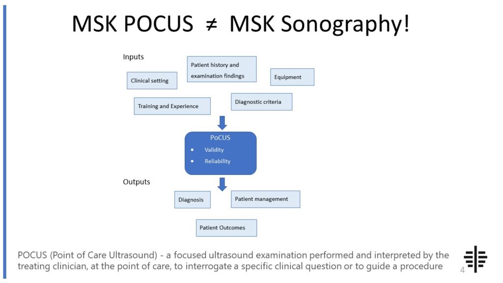
The POCUS process is very different to traditional US based in a radiology establishment. And POCUS practitioners need to be aware of those factors, unique to their particular situation, that influence diagnostic accuracy. That was the topic I presented at the plenary session of the NZAMM Annual Scientific Meeting in Wellington. A picture says 1000 ...

Posted by on 2022-10-04
We’re proud to announce that the New Zealand College of Musculoskeletal Medicine has endorsed our POCUS courses for CME and as part of vocational training. The NZCMM is responsible for setting the high standards and training of Specialist Musculoskeletal Medicine Physicians in New Zealand. NZCMM endorsement is an acknowledgement that our courses meet these standards. ...

Posted by on 2022-06-23
The role of a pressure transducer in measuring blood pressure is crucial. Blood pressure is the force exerted by circulating blood on the walls of blood vessels. A pressure transducer is used to measure this force by converting it into an electrical signal. In the case of blood pressure measurement, the transducer is typically placed inside a cuff or a catheter. When the cuff is inflated or the catheter is inserted into a blood vessel, the pressure exerted by the blood compresses the transducer. This compression causes a change in the electrical resistance or capacitance of the transducer, which is then converted into a measurable signal that represents the blood pressure.

A thermocouple transducer is commonly used to convert temperature into an electrical signal. It consists of two different metal wires, known as thermocouple wires, that are joined together at one end. When there is a temperature difference between the joined end and the other end of the wires, a voltage is generated. This voltage is proportional to the temperature difference and can be measured using a voltmeter. By calibrating the thermocouple transducer with known temperature values, the voltage output can be correlated to the actual temperature being measured, allowing for accurate temperature monitoring in various applications.
Capacitive transducers offer several advantages in pressure sensing applications. One advantage is their high sensitivity, as they can detect even small changes in capacitance. This makes them suitable for measuring low-pressure ranges. Additionally, capacitive transducers are not affected by magnetic fields, which can be a concern in certain environments. They also have a wide frequency response, allowing for accurate measurements across a range of frequencies. Furthermore, capacitive transducers are compact and can be easily integrated into different systems, making them versatile and convenient for pressure sensing applications.

A strain gauge transducer measures the deformation of a material by utilizing the principle of electrical resistance. It consists of a thin wire or foil that is attached to the material being measured. When the material undergoes deformation, the wire or foil also stretches or compresses, causing a change in its electrical resistance. This change in resistance is then measured using a Wheatstone bridge circuit, which converts it into an electrical signal that represents the strain or deformation of the material. Strain gauge transducers are commonly used in applications such as structural monitoring, load cells, and force measurements.
The principle behind the operation of a photoelectric transducer in optical sensing is based on the detection of light and its conversion into an electrical signal. Photoelectric transducers consist of a light-sensitive element, such as a photodiode or a phototransistor, which generates an electrical current when exposed to light. When light falls on the light-sensitive element, it excites electrons, causing them to move and create a flow of current. The intensity of the light determines the magnitude of the current generated. This electrical signal can then be processed and used for various optical sensing applications, such as detecting the presence or absence of objects, measuring distances, or reading barcodes.

Musculoskeletal ultrasound is a valuable tool for assessing sacroiliac joint dysfunction, but it does have some limitations. One limitation is that it can be challenging to obtain clear and accurate images of the sacroiliac joint due to its deep location and the presence of overlying structures such as muscles and ligaments. Additionally, the interpretation of ultrasound images can be subjective and dependent on the experience and expertise of the operator. Another limitation is that ultrasound may not be able to provide a comprehensive assessment of the sacroiliac joint, as it may not be able to visualize certain structures such as the articular cartilage or the joint space. Furthermore, ultrasound is limited in its ability to assess the functional aspects of the sacroiliac joint, such as joint mobility or stability. Therefore, while musculoskeletal ultrasound can be a useful tool in the assessment of sacroiliac joint dysfunction, it should be used in conjunction with other imaging modalities and clinical findings to ensure a comprehensive evaluation.
Musculoskeletal ultrasound can be a useful tool for diagnosing vertebral compression fractures. This imaging technique utilizes high-frequency sound waves to create detailed images of the musculoskeletal system, including the spine. By examining the affected area, musculoskeletal ultrasound can help identify signs of vertebral compression fractures, such as changes in bone density, deformities, or the presence of fractures. Additionally, musculoskeletal ultrasound can provide real-time visualization, allowing for dynamic assessment of the spine during movement or weight-bearing activities. This can aid in the accurate diagnosis and monitoring of vertebral compression fractures, guiding appropriate treatment strategies.
Ultrasound is a commonly used imaging technique for diagnosing musculoskeletal conditions, but it does have its limitations. One limitation is its inability to penetrate bone, which can make it difficult to visualize deep structures or assess fractures. Additionally, ultrasound is operator-dependent, meaning that the quality of the images obtained can vary depending on the skill and experience of the person performing the examination. This can lead to inconsistencies in diagnosis and potentially missed or misinterpreted findings. Another limitation is the limited field of view provided by ultrasound, which may make it challenging to assess larger areas or multiple structures simultaneously. Finally, ultrasound is not always able to provide detailed information about the composition of tissues, such as differentiating between different types of soft tissue masses. In these cases, additional imaging modalities, such as MRI or CT scans, may be necessary for a more comprehensive evaluation.
Musculoskeletal ultrasound has been found to be effective in diagnosing osteoid osteoma. This imaging technique utilizes high-frequency sound waves to create detailed images of the musculoskeletal system, allowing for the visualization of bone and soft tissue structures. By examining the affected area, musculoskeletal ultrasound can detect the characteristic features of osteoid osteoma, such as a central nidus surrounded by reactive bone formation. Additionally, this modality can provide real-time imaging, allowing for dynamic assessment of the lesion and its surrounding structures. The use of musculoskeletal ultrasound in diagnosing osteoid osteoma can help guide treatment decisions and minimize the need for more invasive procedures.
Musculoskeletal ultrasound has been found to be a valuable tool in the diagnosis of osteonecrosis of the femoral head. This imaging technique utilizes high-frequency sound waves to create detailed images of the musculoskeletal system, allowing for the visualization of bone and soft tissue structures. By examining the femoral head using ultrasound, healthcare professionals can identify characteristic findings associated with osteonecrosis, such as subchondral lucency, cortical collapse, and irregularity of the articular surface. Additionally, musculoskeletal ultrasound can provide real-time imaging, allowing for dynamic assessment of the affected area. While other imaging modalities like magnetic resonance imaging (MRI) are considered the gold standard for diagnosing osteonecrosis, musculoskeletal ultrasound can serve as a useful adjunctive tool, particularly in cases where MRI is contraindicated or unavailable. Overall, musculoskeletal ultrasound has demonstrated effectiveness in diagnosing osteonecrosis of the femoral head, providing valuable information for treatment planning and management.