
The accuracy of ultrasound in detecting fractures can vary depending on the type and location of the fracture. In general, ultrasound has been found to be highly accurate in detecting fractures of the extremities, such as the wrist, ankle, and fingers. Studies have shown that ultrasound has a sensitivity ranging from 80% to 100% in detecting fractures in these areas. However, the accuracy of ultrasound in detecting fractures in other areas, such as the spine or pelvis, may be lower and may require additional imaging techniques for confirmation.
There are several advantages of using ultrasound for fracture evaluation compared to other imaging techniques. Firstly, ultrasound is a non-invasive and radiation-free imaging modality, making it a safer option, especially for children and pregnant women. Secondly, ultrasound can provide real-time imaging, allowing for dynamic evaluation of the fracture and surrounding structures. This can be particularly useful in assessing joint stability and detecting any associated soft tissue injuries. Additionally, ultrasound is readily available, portable, and relatively cost-effective compared to other imaging modalities, making it a convenient option for fracture evaluation.
Over the last couple of years, we’ve brought you several courses focusing on Ultrasound Guided Injection Techniques. They’ve been extremely popular, and like our other courses, the feedback has been fantastic. One thing we’ve learnt along the way is that to get the most out of learning injection techniques, a solid grounding in MSK Ultrasound ...
Posted by on 2024-02-10
What a year 2023 was! We’ve loved bringing you courses covering US of the upper and lower limb, and US guided injections through the year. The mix of health professionals from all sorts of backgrounds (Doctors, Nurses, Physios, Sonographers to name a few) has been amazing to be part of. We’ve been humbled by your ...
Posted by on 2023-09-17
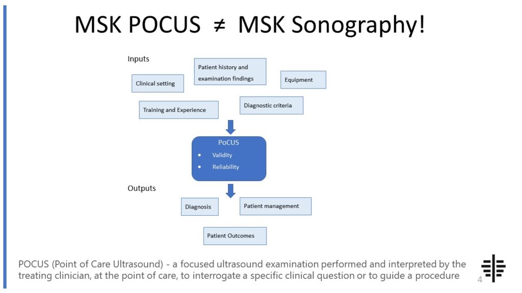
The POCUS process is very different to traditional US based in a radiology establishment. And POCUS practitioners need to be aware of those factors, unique to their particular situation, that influence diagnostic accuracy. That was the topic I presented at the plenary session of the NZAMM Annual Scientific Meeting in Wellington. A picture says 1000 ...

Posted by on 2022-10-04
We’re proud to announce that the New Zealand College of Musculoskeletal Medicine has endorsed our POCUS courses for CME and as part of vocational training. The NZCMM is responsible for setting the high standards and training of Specialist Musculoskeletal Medicine Physicians in New Zealand. NZCMM endorsement is an acknowledgement that our courses meet these standards. ...

Posted by on 2022-06-23
Ultrasound can be used to evaluate many types of fractures, particularly those involving the extremities. Fractures of the wrist, ankle, fingers, and toes are commonly evaluated using ultrasound. However, the utility of ultrasound in evaluating fractures in other areas, such as the spine or pelvis, may be limited due to the presence of overlying structures that can hinder visualization. In these cases, other imaging techniques, such as X-ray, CT scan, or MRI, may be necessary for a more comprehensive evaluation.

Despite its advantages, there are some limitations and drawbacks to using ultrasound for fracture evaluation. One limitation is the operator-dependency of ultrasound, as the quality of the images obtained can vary based on the skill and experience of the operator. Additionally, ultrasound may not be able to provide detailed information about the bony anatomy or the extent of the fracture, especially in complex or comminuted fractures. In these cases, additional imaging modalities may be required for a more accurate assessment.
Ultrasound can help in determining the severity of a fracture by assessing various factors. Firstly, ultrasound can visualize the displacement or angulation of the fracture fragments, which can indicate the degree of instability and potential for complications. Secondly, ultrasound can assess the integrity of the surrounding soft tissues, such as tendons, ligaments, and muscles, which can be affected by the fracture. Lastly, ultrasound can evaluate the blood flow in the area, which can provide information about the vascularity and healing potential of the fracture.

Yes, ultrasound can be used to monitor the healing process of a fracture. During follow-up appointments, ultrasound can assess the progression of bone healing by visualizing the formation of callus, which is the new bone that forms during the healing process. Ultrasound can also evaluate the surrounding soft tissues for any signs of inflammation or complications, such as infection or delayed healing. Regular ultrasound examinations can help track the healing progress and guide the management of the fracture.
While there are no specific protocols or guidelines for performing ultrasound for fracture evaluation, there are general principles that should be followed. It is important to use high-frequency transducers to achieve better resolution and visualization of the fracture site. The examination should include both longitudinal and transverse views to obtain a comprehensive assessment. The operator should have a good understanding of the anatomy and biomechanics of the area being evaluated to accurately identify and characterize the fracture. Additionally, it is important to compare the ultrasound findings with clinical history, physical examination, and other imaging modalities, if available, to ensure an accurate diagnosis and appropriate management.

Musculoskeletal ultrasound plays a crucial role in diagnosing osteochondral lesions by providing detailed imaging of the affected area. This imaging technique utilizes high-frequency sound waves to create real-time images of the musculoskeletal system, allowing healthcare professionals to visualize the structure and integrity of the bones, cartilage, and surrounding soft tissues. By using musculoskeletal ultrasound, clinicians can accurately assess the size, location, and severity of osteochondral lesions, as well as identify any associated abnormalities such as bone spurs or joint effusion. Additionally, this imaging modality enables dynamic evaluation of joint movement and can help differentiate between acute and chronic lesions. Overall, musculoskeletal ultrasound offers a non-invasive and cost-effective method for diagnosing osteochondral lesions, aiding in the development of appropriate treatment plans and improving patient outcomes.
Assessing spinal stenosis using musculoskeletal ultrasound presents several challenges. Firstly, the limited penetration depth of ultrasound waves may hinder the visualization of deep structures within the spine, particularly in patients with a high body mass index or those with excessive subcutaneous fat. Additionally, the complex anatomy of the spine, with its multiple layers of muscles, ligaments, and bones, can make it difficult to accurately identify and assess the extent of stenosis using ultrasound alone. Furthermore, the dynamic nature of spinal stenosis, which can worsen or improve with changes in posture or movement, may require real-time imaging techniques that ultrasound may not be able to provide. Lastly, the operator's expertise and experience in performing musculoskeletal ultrasound for spinal stenosis assessment is crucial, as the interpretation of ultrasound images can be subjective and require a deep understanding of spinal anatomy and pathology.
Musculoskeletal ultrasound has the potential to identify ligamentous laxity in patients with joint hypermobility syndrome. By utilizing this imaging technique, healthcare professionals can visualize the ligaments and assess their integrity and stability. The ultrasound can detect any abnormalities or laxity in the ligaments, providing valuable information about the extent of joint instability in individuals with joint hypermobility syndrome. Additionally, musculoskeletal ultrasound can also help in evaluating the surrounding structures such as tendons and muscles, which may contribute to joint instability. This comprehensive assessment can aid in the diagnosis and management of joint hypermobility syndrome, allowing for targeted treatment strategies to improve patient outcomes.
Musculoskeletal ultrasound plays a crucial role in the diagnosis of tendon injuries by providing detailed imaging of the affected area. This non-invasive imaging technique utilizes high-frequency sound waves to create real-time images of the tendons, allowing healthcare professionals to assess the integrity and identify any abnormalities or damage. By visualizing the tendon structure, musculoskeletal ultrasound enables the detection of tendon tears, tendinitis, tendinosis, and other tendon-related pathologies. Additionally, this imaging modality allows for dynamic evaluation of the tendons during movement, providing valuable information about tendon function and potential areas of weakness or instability. Overall, musculoskeletal ultrasound aids in the accurate diagnosis of tendon injuries, guiding appropriate treatment strategies and facilitating optimal patient care.
Musculoskeletal ultrasound plays a crucial role in diagnosing stress injuries in athletes by providing detailed imaging of the musculoskeletal system. This non-invasive imaging technique utilizes high-frequency sound waves to create real-time images of the bones, muscles, tendons, and ligaments. By examining these structures, healthcare professionals can identify any abnormalities or damage that may be indicative of a stress injury. Musculoskeletal ultrasound allows for the visualization of stress fractures, muscle tears, tendonitis, and other soft tissue injuries, providing valuable information for accurate diagnosis and treatment planning. Additionally, this imaging modality enables dynamic assessment, allowing healthcare professionals to evaluate the affected area during movement or stress, which can further aid in the diagnosis of stress injuries in athletes. Overall, musculoskeletal ultrasound is a valuable tool in the diagnostic process, helping healthcare professionals effectively identify and manage stress injuries in athletes.